Chlorine »
PDB 7z70-7zkr »
7ziz »
Chlorine in PDB 7ziz: X-Ray Structure of the Dead Variant Haloalkane Dehalogenase HALOTAG7- D106A Bound to A Pentanol Tetramethylrhodamine Ligand (Tmr-HY5)
Enzymatic activity of X-Ray Structure of the Dead Variant Haloalkane Dehalogenase HALOTAG7- D106A Bound to A Pentanol Tetramethylrhodamine Ligand (Tmr-HY5)
All present enzymatic activity of X-Ray Structure of the Dead Variant Haloalkane Dehalogenase HALOTAG7- D106A Bound to A Pentanol Tetramethylrhodamine Ligand (Tmr-HY5):
3.8.1.5;
3.8.1.5;
Protein crystallography data
The structure of X-Ray Structure of the Dead Variant Haloalkane Dehalogenase HALOTAG7- D106A Bound to A Pentanol Tetramethylrhodamine Ligand (Tmr-HY5), PDB code: 7ziz
was solved by
M.Tarnawski,
J.Kompa,
K.Johnsson,
J.Hiblot,
with X-Ray Crystallography technique. A brief refinement statistics is given in the table below:
| Resolution Low / High (Å) | 44.35 / 1.50 |
| Space group | P 21 21 2 |
| Cell size a, b, c (Å), α, β, γ (°) | 77.83, 88.71, 44.2, 90, 90, 90 |
| R / Rfree (%) | 16.9 / 19.5 |
Chlorine Binding Sites:
The binding sites of Chlorine atom in the X-Ray Structure of the Dead Variant Haloalkane Dehalogenase HALOTAG7- D106A Bound to A Pentanol Tetramethylrhodamine Ligand (Tmr-HY5)
(pdb code 7ziz). This binding sites where shown within
5.0 Angstroms radius around Chlorine atom.
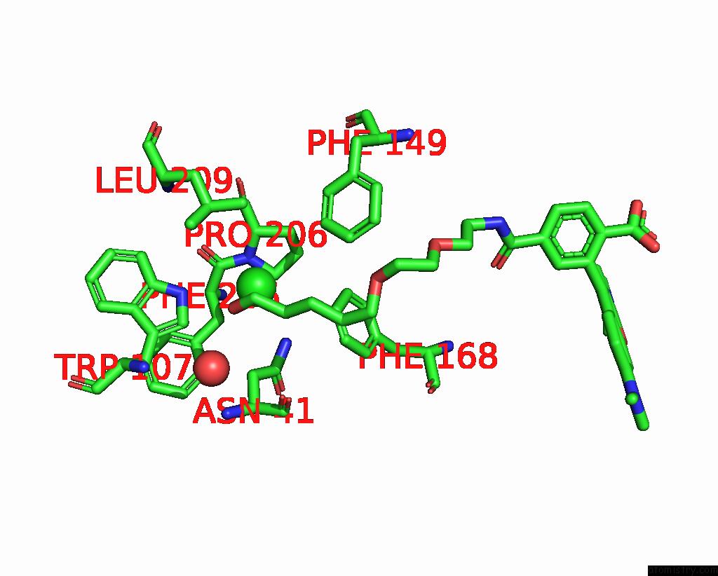
In total only one binding site of Chlorine was determined in the X-Ray Structure of the Dead Variant Haloalkane Dehalogenase HALOTAG7- D106A Bound to A Pentanol Tetramethylrhodamine Ligand (Tmr-HY5), PDB code: 7ziz:
In total only one binding site of Chlorine was determined in the X-Ray Structure of the Dead Variant Haloalkane Dehalogenase HALOTAG7- D106A Bound to A Pentanol Tetramethylrhodamine Ligand (Tmr-HY5), PDB code: 7ziz:
Chlorine binding site 1 out of 1 in 7ziz
Go back to
Chlorine binding site 1 out
of 1 in the X-Ray Structure of the Dead Variant Haloalkane Dehalogenase HALOTAG7- D106A Bound to A Pentanol Tetramethylrhodamine Ligand (Tmr-HY5)
Mono view
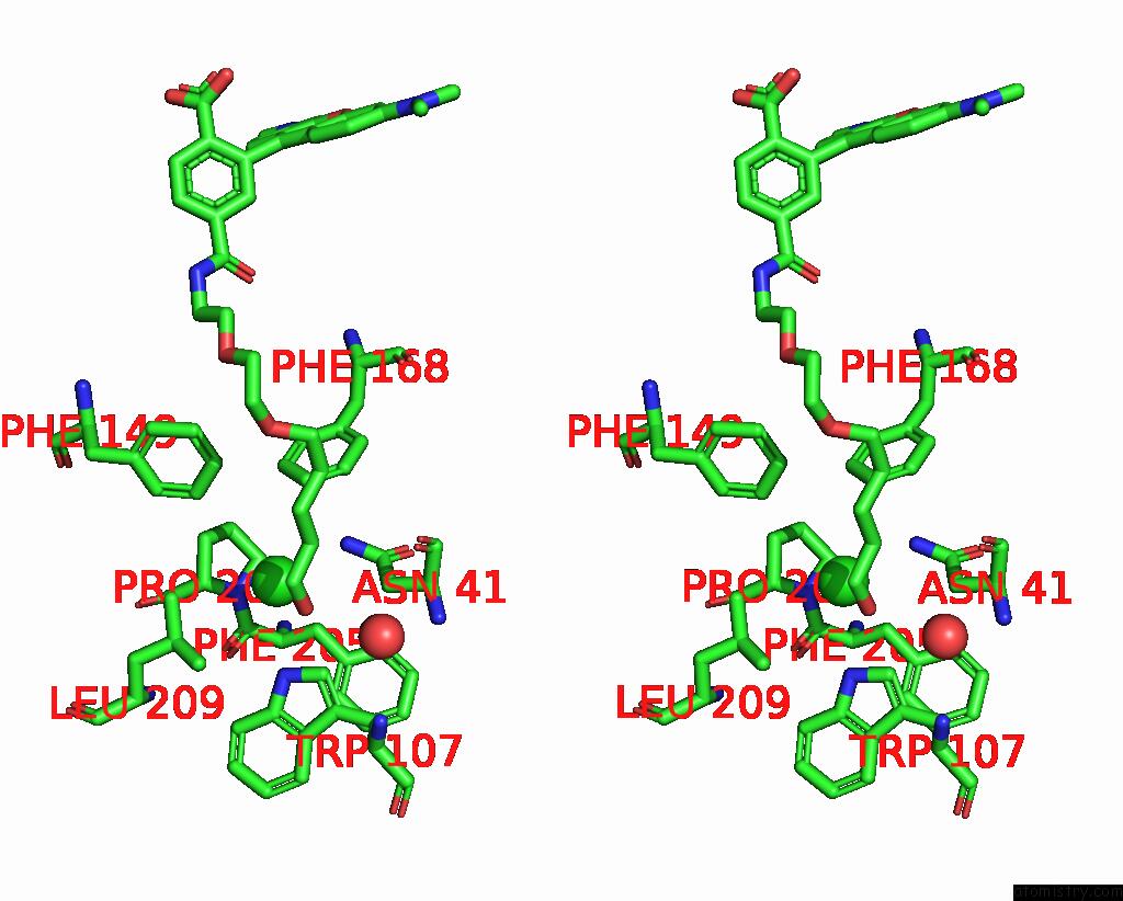
Stereo pair view
Mono view
Stereo pair view
A full contact list of Chlorine with other atoms in the Cl binding
site number 1 of X-Ray Structure of the Dead Variant Haloalkane Dehalogenase HALOTAG7- D106A Bound to A Pentanol Tetramethylrhodamine Ligand (Tmr-HY5) within 5.0Å range:
|
Reference:
J.Kompa,
J.Bruins,
M.Glogger,
J.Wilhelm,
M.S.Frei,
M.Tarnawski,
E.D’Este,
M.Heilemann,
J.Hiblot,
K.Johnsson.
Exchangeable Halotag Ligands For Super-Resolution Fluorescence Microscopy J.Am.Chem.Soc. 2023.
ISSN: ESSN 1520-5126
DOI: 10.1021/JACS.2C11969
Page generated: Tue Jul 30 06:15:44 2024
ISSN: ESSN 1520-5126
DOI: 10.1021/JACS.2C11969
Last articles
Zn in 9JYWZn in 9IR4
Zn in 9IR3
Zn in 9GMX
Zn in 9GMW
Zn in 9JEJ
Zn in 9ERF
Zn in 9ERE
Zn in 9EGV
Zn in 9EGW