Chlorine »
PDB 7zwy-8a2c »
8a1o »
Chlorine in PDB 8a1o: Crystal Structure of the Transpeptidase LDTMT2 From Mycobacterium Tuberculosis in Complex with Acrylamide Analogue 8
Protein crystallography data
The structure of Crystal Structure of the Transpeptidase LDTMT2 From Mycobacterium Tuberculosis in Complex with Acrylamide Analogue 8, PDB code: 8a1o
was solved by
M.De Munnik,
P.A.Lang,
J.Brem,
C.J.Schofield,
with X-Ray Crystallography technique. A brief refinement statistics is given in the table below:
| Resolution Low / High (Å) | 46.50 / 1.95 |
| Space group | P 21 21 2 |
| Cell size a, b, c (Å), α, β, γ (°) | 76.87, 93, 61.4, 90, 90, 90 |
| R / Rfree (%) | 18.8 / 22.5 |
Other elements in 8a1o:
The structure of Crystal Structure of the Transpeptidase LDTMT2 From Mycobacterium Tuberculosis in Complex with Acrylamide Analogue 8 also contains other interesting chemical elements:
| Sodium | (Na) | 1 atom |
Chlorine Binding Sites:
The binding sites of Chlorine atom in the Crystal Structure of the Transpeptidase LDTMT2 From Mycobacterium Tuberculosis in Complex with Acrylamide Analogue 8
(pdb code 8a1o). This binding sites where shown within
5.0 Angstroms radius around Chlorine atom.
In total 4 binding sites of Chlorine where determined in the Crystal Structure of the Transpeptidase LDTMT2 From Mycobacterium Tuberculosis in Complex with Acrylamide Analogue 8, PDB code: 8a1o:
Jump to Chlorine binding site number: 1; 2; 3; 4;
In total 4 binding sites of Chlorine where determined in the Crystal Structure of the Transpeptidase LDTMT2 From Mycobacterium Tuberculosis in Complex with Acrylamide Analogue 8, PDB code: 8a1o:
Jump to Chlorine binding site number: 1; 2; 3; 4;
Chlorine binding site 1 out of 4 in 8a1o
Go back to
Chlorine binding site 1 out
of 4 in the Crystal Structure of the Transpeptidase LDTMT2 From Mycobacterium Tuberculosis in Complex with Acrylamide Analogue 8

Mono view
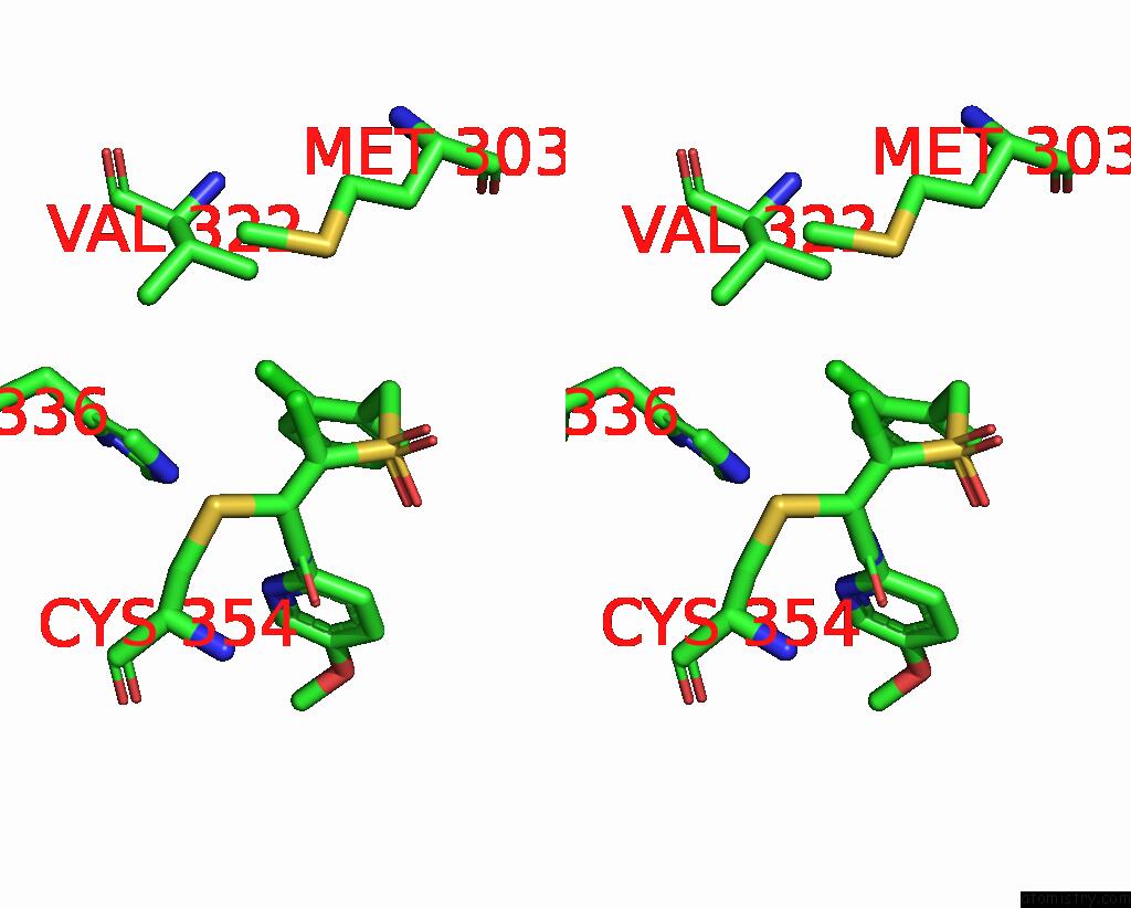
Stereo pair view
Mono view
Stereo pair view
A full contact list of Chlorine with other atoms in the Cl binding
site number 1 of Crystal Structure of the Transpeptidase LDTMT2 From Mycobacterium Tuberculosis in Complex with Acrylamide Analogue 8 within 5.0Å range:
|
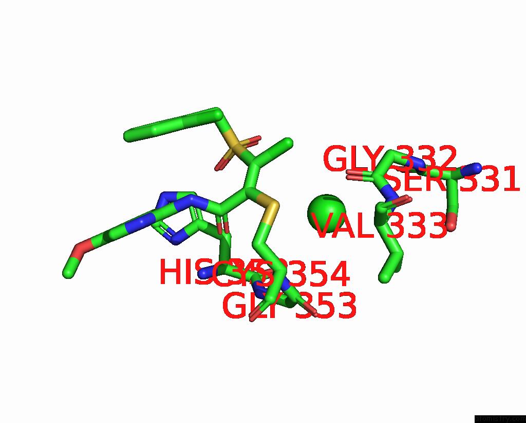
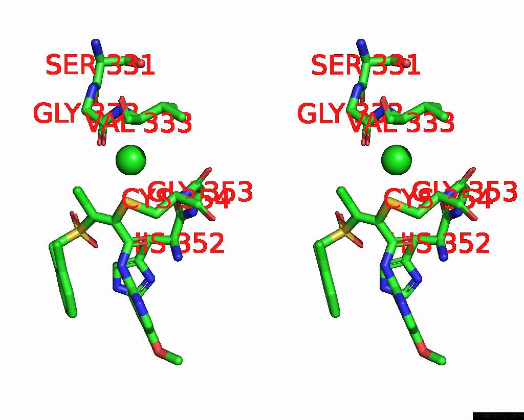
Chlorine binding site 2 out of 4 in 8a1o
Go back to
Chlorine binding site 2 out
of 4 in the Crystal Structure of the Transpeptidase LDTMT2 From Mycobacterium Tuberculosis in Complex with Acrylamide Analogue 8

Mono view

Stereo pair view
Mono view
Stereo pair view
A full contact list of Chlorine with other atoms in the Cl binding
site number 2 of Crystal Structure of the Transpeptidase LDTMT2 From Mycobacterium Tuberculosis in Complex with Acrylamide Analogue 8 within 5.0Å range:
|
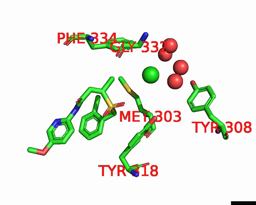
Chlorine binding site 3 out of 4 in 8a1o
Go back to
Chlorine binding site 3 out
of 4 in the Crystal Structure of the Transpeptidase LDTMT2 From Mycobacterium Tuberculosis in Complex with Acrylamide Analogue 8
Mono view
Stereo pair view
Mono view
Stereo pair view
A full contact list of Chlorine with other atoms in the Cl binding
site number 3 of Crystal Structure of the Transpeptidase LDTMT2 From Mycobacterium Tuberculosis in Complex with Acrylamide Analogue 8 within 5.0Å range:
|
Chlorine binding site 4 out of 4 in 8a1o
Go back to
Chlorine binding site 4 out
of 4 in the Crystal Structure of the Transpeptidase LDTMT2 From Mycobacterium Tuberculosis in Complex with Acrylamide Analogue 8
Mono view

Stereo pair view
Mono view
Stereo pair view
A full contact list of Chlorine with other atoms in the Cl binding
site number 4 of Crystal Structure of the Transpeptidase LDTMT2 From Mycobacterium Tuberculosis in Complex with Acrylamide Analogue 8 within 5.0Å range:
|
Reference:
M.De Munnik,
P.A.Lang,
J.Brem,
C.J.Schofield.
Crystal Structure of the Transpeptidase LDTMT2 From Mycobacterium Tuberculosis in Complex with Acrylamide Analogue 8 To Be Published.
Page generated: Tue Jul 30 06:27:54 2024
Last articles
Zn in 9JYWZn in 9IR4
Zn in 9IR3
Zn in 9GMX
Zn in 9GMW
Zn in 9JEJ
Zn in 9ERF
Zn in 9ERE
Zn in 9EGV
Zn in 9EGW