Chlorine »
PDB 8a19-8aau »
8a1z »
Chlorine in PDB 8a1z: Crystal Structure of Phosphoserine Phosphatase Serb From Mycobacterium Avium in Complex with 1-(2,4-Dichlorophenyl)-3-Hydroxyurea
Enzymatic activity of Crystal Structure of Phosphoserine Phosphatase Serb From Mycobacterium Avium in Complex with 1-(2,4-Dichlorophenyl)-3-Hydroxyurea
All present enzymatic activity of Crystal Structure of Phosphoserine Phosphatase Serb From Mycobacterium Avium in Complex with 1-(2,4-Dichlorophenyl)-3-Hydroxyurea:
3.1.3.3;
3.1.3.3;
Protein crystallography data
The structure of Crystal Structure of Phosphoserine Phosphatase Serb From Mycobacterium Avium in Complex with 1-(2,4-Dichlorophenyl)-3-Hydroxyurea, PDB code: 8a1z
was solved by
M.Haufroid,
J.Wouters,
with X-Ray Crystallography technique. A brief refinement statistics is given in the table below:
| Resolution Low / High (Å) | 43.59 / 2.28 |
| Space group | I 2 2 2 |
| Cell size a, b, c (Å), α, β, γ (°) | 68.6, 107.7, 132.6, 90, 90, 90 |
| R / Rfree (%) | 20.6 / 24.6 |
Other elements in 8a1z:
The structure of Crystal Structure of Phosphoserine Phosphatase Serb From Mycobacterium Avium in Complex with 1-(2,4-Dichlorophenyl)-3-Hydroxyurea also contains other interesting chemical elements:
| Magnesium | (Mg) | 1 atom |
Chlorine Binding Sites:
The binding sites of Chlorine atom in the Crystal Structure of Phosphoserine Phosphatase Serb From Mycobacterium Avium in Complex with 1-(2,4-Dichlorophenyl)-3-Hydroxyurea
(pdb code 8a1z). This binding sites where shown within
5.0 Angstroms radius around Chlorine atom.
In total 3 binding sites of Chlorine where determined in the Crystal Structure of Phosphoserine Phosphatase Serb From Mycobacterium Avium in Complex with 1-(2,4-Dichlorophenyl)-3-Hydroxyurea, PDB code: 8a1z:
Jump to Chlorine binding site number: 1; 2; 3;
In total 3 binding sites of Chlorine where determined in the Crystal Structure of Phosphoserine Phosphatase Serb From Mycobacterium Avium in Complex with 1-(2,4-Dichlorophenyl)-3-Hydroxyurea, PDB code: 8a1z:
Jump to Chlorine binding site number: 1; 2; 3;
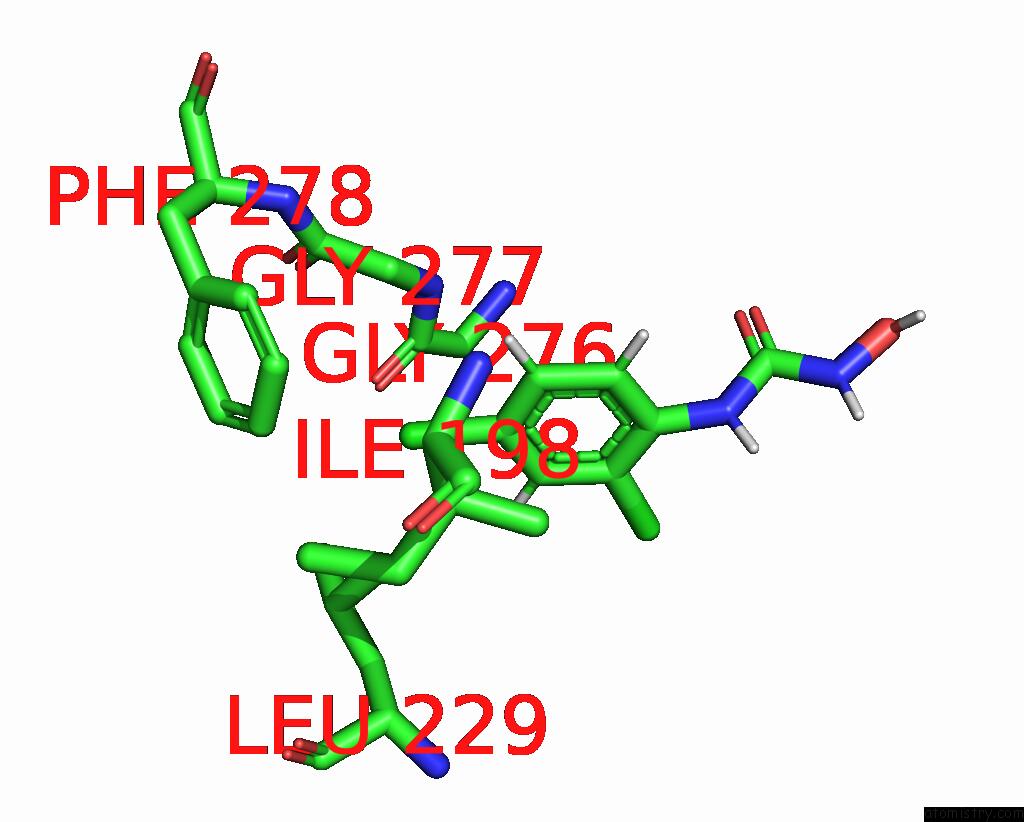
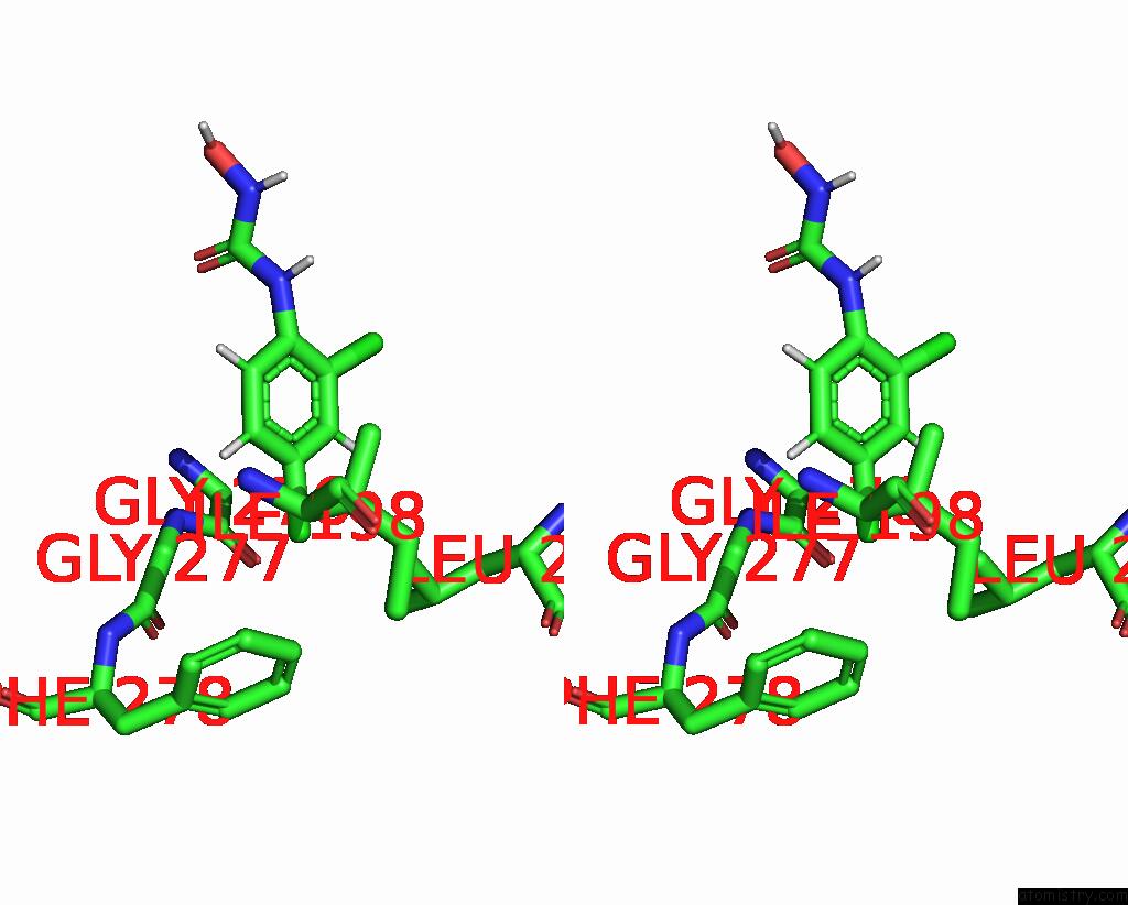
Chlorine binding site 1 out of 3 in 8a1z
Go back to
Chlorine binding site 1 out
of 3 in the Crystal Structure of Phosphoserine Phosphatase Serb From Mycobacterium Avium in Complex with 1-(2,4-Dichlorophenyl)-3-Hydroxyurea

Mono view

Stereo pair view
Mono view
Stereo pair view
A full contact list of Chlorine with other atoms in the Cl binding
site number 1 of Crystal Structure of Phosphoserine Phosphatase Serb From Mycobacterium Avium in Complex with 1-(2,4-Dichlorophenyl)-3-Hydroxyurea within 5.0Å range:
|
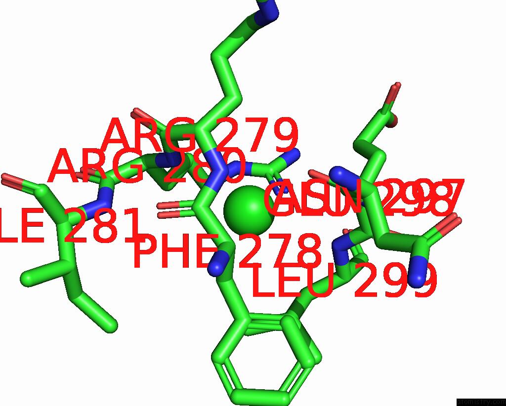
Chlorine binding site 2 out of 3 in 8a1z
Go back to
Chlorine binding site 2 out
of 3 in the Crystal Structure of Phosphoserine Phosphatase Serb From Mycobacterium Avium in Complex with 1-(2,4-Dichlorophenyl)-3-Hydroxyurea
Mono view
Stereo pair view
Mono view
Stereo pair view
A full contact list of Chlorine with other atoms in the Cl binding
site number 2 of Crystal Structure of Phosphoserine Phosphatase Serb From Mycobacterium Avium in Complex with 1-(2,4-Dichlorophenyl)-3-Hydroxyurea within 5.0Å range:
|
Chlorine binding site 3 out of 3 in 8a1z
Go back to
Chlorine binding site 3 out
of 3 in the Crystal Structure of Phosphoserine Phosphatase Serb From Mycobacterium Avium in Complex with 1-(2,4-Dichlorophenyl)-3-Hydroxyurea
Mono view

Stereo pair view
Mono view
Stereo pair view
A full contact list of Chlorine with other atoms in the Cl binding
site number 3 of Crystal Structure of Phosphoserine Phosphatase Serb From Mycobacterium Avium in Complex with 1-(2,4-Dichlorophenyl)-3-Hydroxyurea within 5.0Å range:
|
Reference:
M.Haufroid,
A.N.Volkov,
J.Wouters.
Targeting the Phosphoserine Phosphatase MTSERB2 For Tuberculosis Drug Discovery, An Hybrid Knowledge Based /Fragment Based Approach. Eur.J.Med.Chem. V. 245 14935 2022.
ISSN: ISSN 0223-5234
PubMed: 36403421
DOI: 10.1016/J.EJMECH.2022.114935
Page generated: Sun Jul 13 08:57:56 2025
ISSN: ISSN 0223-5234
PubMed: 36403421
DOI: 10.1016/J.EJMECH.2022.114935
Last articles
Fe in 2HREFe in 2HV4
Fe in 2HU9
Fe in 2HUO
Fe in 2HT9
Fe in 2HRC
Fe in 2HR5
Fe in 2HMZ
Fe in 2HMQ
Fe in 2HPD