Chlorine »
PDB 8ab7-8alv »
8ak7 »
Chlorine in PDB 8ak7: Human SIRT6 in Complex with Adp-Ribose and Fragment 6-O-Methylguanine
Enzymatic activity of Human SIRT6 in Complex with Adp-Ribose and Fragment 6-O-Methylguanine
All present enzymatic activity of Human SIRT6 in Complex with Adp-Ribose and Fragment 6-O-Methylguanine:
2.3.1.286;
2.3.1.286;
Protein crystallography data
The structure of Human SIRT6 in Complex with Adp-Ribose and Fragment 6-O-Methylguanine, PDB code: 8ak7
was solved by
W.You,
C.Steegborn,
with X-Ray Crystallography technique. A brief refinement statistics is given in the table below:
| Resolution Low / High (Å) | 43.40 / 1.81 |
| Space group | P 63 |
| Cell size a, b, c (Å), α, β, γ (°) | 91.072, 91.072, 143.251, 90, 90, 120 |
| R / Rfree (%) | 20.1 / 23.3 |
Other elements in 8ak7:
The structure of Human SIRT6 in Complex with Adp-Ribose and Fragment 6-O-Methylguanine also contains other interesting chemical elements:
| Zinc | (Zn) | 2 atoms |
Chlorine Binding Sites:
The binding sites of Chlorine atom in the Human SIRT6 in Complex with Adp-Ribose and Fragment 6-O-Methylguanine
(pdb code 8ak7). This binding sites where shown within
5.0 Angstroms radius around Chlorine atom.
In total 4 binding sites of Chlorine where determined in the Human SIRT6 in Complex with Adp-Ribose and Fragment 6-O-Methylguanine, PDB code: 8ak7:
Jump to Chlorine binding site number: 1; 2; 3; 4;
In total 4 binding sites of Chlorine where determined in the Human SIRT6 in Complex with Adp-Ribose and Fragment 6-O-Methylguanine, PDB code: 8ak7:
Jump to Chlorine binding site number: 1; 2; 3; 4;
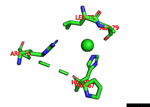
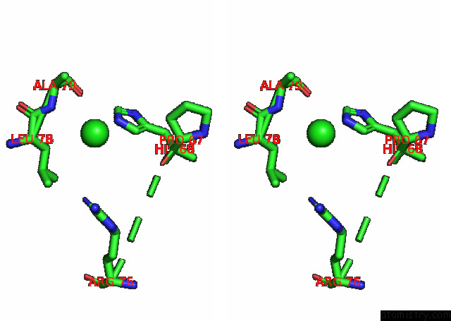
Chlorine binding site 1 out of 4 in 8ak7
Go back to
Chlorine binding site 1 out
of 4 in the Human SIRT6 in Complex with Adp-Ribose and Fragment 6-O-Methylguanine
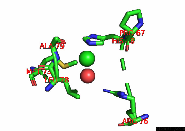
Mono view
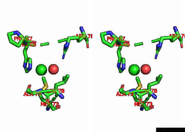
Stereo pair view
Mono view
Stereo pair view
A full contact list of Chlorine with other atoms in the Cl binding
site number 1 of Human SIRT6 in Complex with Adp-Ribose and Fragment 6-O-Methylguanine within 5.0Å range:
|
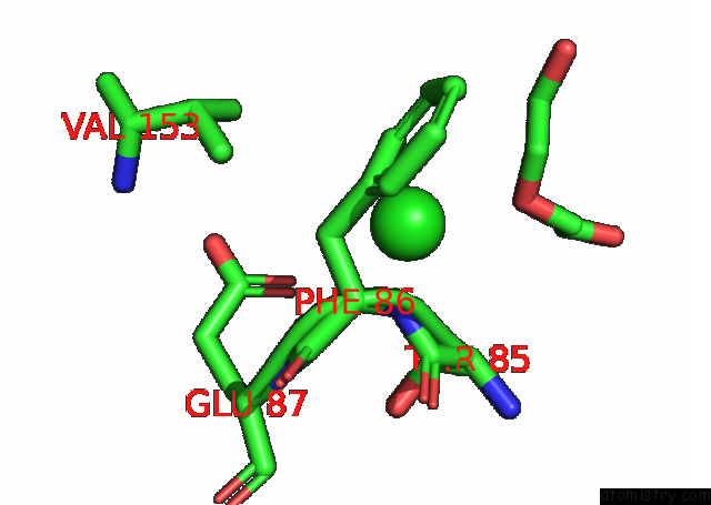
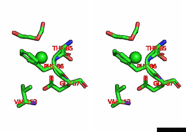
Chlorine binding site 2 out of 4 in 8ak7
Go back to
Chlorine binding site 2 out
of 4 in the Human SIRT6 in Complex with Adp-Ribose and Fragment 6-O-Methylguanine
Mono view
Stereo pair view
Mono view
Stereo pair view
A full contact list of Chlorine with other atoms in the Cl binding
site number 2 of Human SIRT6 in Complex with Adp-Ribose and Fragment 6-O-Methylguanine within 5.0Å range:
|
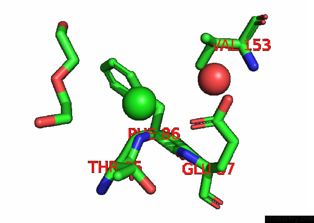
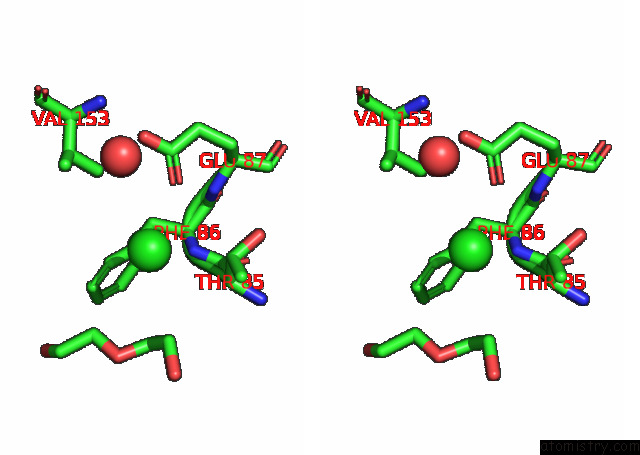
Chlorine binding site 3 out of 4 in 8ak7
Go back to
Chlorine binding site 3 out
of 4 in the Human SIRT6 in Complex with Adp-Ribose and Fragment 6-O-Methylguanine
Mono view
Stereo pair view
Mono view
Stereo pair view
A full contact list of Chlorine with other atoms in the Cl binding
site number 3 of Human SIRT6 in Complex with Adp-Ribose and Fragment 6-O-Methylguanine within 5.0Å range:
|
Chlorine binding site 4 out of 4 in 8ak7
Go back to
Chlorine binding site 4 out
of 4 in the Human SIRT6 in Complex with Adp-Ribose and Fragment 6-O-Methylguanine
Mono view
Stereo pair view
Mono view
Stereo pair view
A full contact list of Chlorine with other atoms in the Cl binding
site number 4 of Human SIRT6 in Complex with Adp-Ribose and Fragment 6-O-Methylguanine within 5.0Å range:
|
Reference:
W.You,
C.Steegborn.
Development of Novel Sirtuin 6 Inhibitors and Activators Based on A Protein Crystallography-Based Fragment Screen To Be Published.
Page generated: Sun Jul 13 09:08:05 2025
Last articles
F in 7NOSF in 7NP5
F in 7NDV
F in 7NP6
F in 7NOR
F in 7NNB
F in 7NKX
F in 7NLZ
F in 7NLX
F in 7NJ6