Chlorine in PDB 8buc: Structure of DDB1 Bound to DCEMM3-Engaged CDK12-Cyclin K
Enzymatic activity of Structure of DDB1 Bound to DCEMM3-Engaged CDK12-Cyclin K
All present enzymatic activity of Structure of DDB1 Bound to DCEMM3-Engaged CDK12-Cyclin K:
2.7.11.22;
2.7.11.23;
Protein crystallography data
The structure of Structure of DDB1 Bound to DCEMM3-Engaged CDK12-Cyclin K, PDB code: 8buc
was solved by
Z.Kozicka,
G.Kempf,
V.Focht,
N.H.Thoma,
with X-Ray Crystallography technique. A brief refinement statistics is given in the table below:
Resolution Low / High (Å)
|
76.78 /
3.85
|
Space group
|
P 31 2 1
|
Cell size a, b, c (Å), α, β, γ (°)
|
249.773,
249.773,
218.027,
90,
90,
120
|
R / Rfree (%)
|
18.1 /
22.4
|
Chlorine Binding Sites:
The binding sites of Chlorine atom in the Structure of DDB1 Bound to DCEMM3-Engaged CDK12-Cyclin K
(pdb code 8buc). This binding sites where shown within
5.0 Angstroms radius around Chlorine atom.
In total 3 binding sites of Chlorine where determined in the
Structure of DDB1 Bound to DCEMM3-Engaged CDK12-Cyclin K, PDB code: 8buc:
Jump to Chlorine binding site number:
1;
2;
3;
Chlorine binding site 1 out
of 3 in 8buc
Go back to
Chlorine Binding Sites List in 8buc
Chlorine binding site 1 out
of 3 in the Structure of DDB1 Bound to DCEMM3-Engaged CDK12-Cyclin K
Mono view
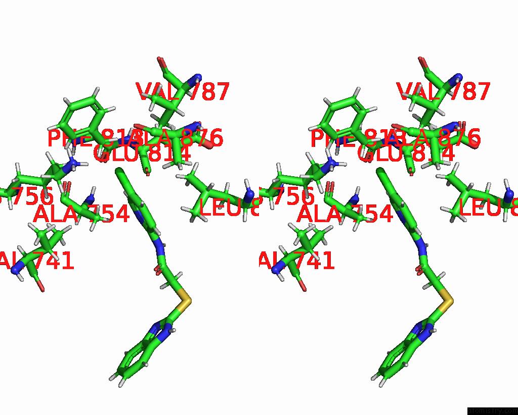
Stereo pair view
|
A full contact list of Chlorine with other atoms in the Cl binding
site number 1 of Structure of DDB1 Bound to DCEMM3-Engaged CDK12-Cyclin K within 5.0Å range:
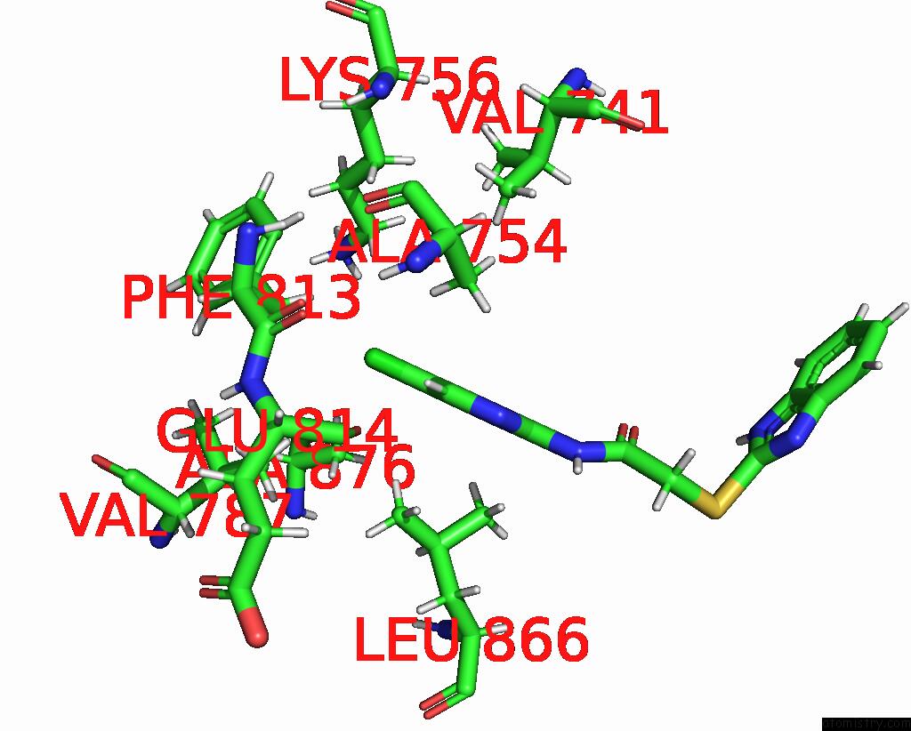
probe
|
atom
|
residue
|
distance (Å)
|
B
|
Occ
|
B:Cl1101
b:125.3
occ:1.00
|
CL1
|
B:RKO1101
|
0.0
|
125.3
|
1.0
|
C4
|
B:RKO1101
|
1.7
|
104.3
|
1.0
|
C3
|
B:RKO1101
|
2.7
|
115.9
|
1.0
|
C5
|
B:RKO1101
|
2.7
|
116.8
|
1.0
|
H2
|
B:RKO1101
|
2.8
|
139.1
|
0.0
|
H3
|
B:RKO1101
|
2.8
|
140.1
|
0.0
|
HB2
|
B:PHE813
|
3.0
|
133.3
|
0.0
|
HB3
|
B:PHE813
|
3.3
|
133.3
|
0.0
|
HG21
|
B:VAL787
|
3.4
|
130.2
|
0.0
|
HD2
|
B:LYS756
|
3.4
|
155.2
|
0.0
|
CB
|
B:PHE813
|
3.4
|
111.1
|
1.0
|
CG
|
B:PHE813
|
3.5
|
118.9
|
1.0
|
CD1
|
B:PHE813
|
3.9
|
113.1
|
1.0
|
HD11
|
B:LEU866
|
3.9
|
126.5
|
0.0
|
N2
|
B:RKO1101
|
4.0
|
124.0
|
1.0
|
HD1
|
B:PHE813
|
4.0
|
135.8
|
0.0
|
C6
|
B:RKO1101
|
4.0
|
107.5
|
1.0
|
HB3
|
B:ALA754
|
4.0
|
149.0
|
0.0
|
CD2
|
B:PHE813
|
4.1
|
137.6
|
1.0
|
HZ2
|
B:LYS756
|
4.2
|
164.2
|
0.0
|
CG2
|
B:VAL787
|
4.3
|
108.5
|
1.0
|
HD2
|
B:PHE813
|
4.3
|
165.1
|
0.0
|
CD
|
B:LYS756
|
4.4
|
129.3
|
1.0
|
HD13
|
B:LEU866
|
4.4
|
126.5
|
0.0
|
HG22
|
B:VAL787
|
4.4
|
130.2
|
0.0
|
O
|
B:GLU814
|
4.4
|
139.9
|
1.0
|
HG3
|
B:LYS756
|
4.4
|
154.7
|
0.0
|
C2
|
B:RKO1101
|
4.4
|
116.7
|
1.0
|
CD1
|
B:LEU866
|
4.6
|
105.4
|
1.0
|
HB1
|
B:ALA876
|
4.6
|
190.0
|
0.0
|
CE1
|
B:PHE813
|
4.7
|
118.2
|
1.0
|
HG11
|
B:VAL787
|
4.7
|
147.6
|
0.0
|
HG23
|
B:VAL787
|
4.7
|
130.2
|
0.0
|
HG11
|
B:VAL741
|
4.7
|
144.8
|
0.0
|
H4
|
B:RKO1101
|
4.7
|
129.1
|
0.0
|
CE2
|
B:PHE813
|
4.8
|
139.0
|
1.0
|
HE3
|
B:LYS756
|
4.8
|
169.7
|
0.0
|
CG
|
B:LYS756
|
4.9
|
128.9
|
1.0
|
CA
|
B:PHE813
|
4.9
|
109.0
|
1.0
|
NZ
|
B:LYS756
|
4.9
|
136.8
|
1.0
|
HG2
|
B:LYS756
|
4.9
|
154.7
|
0.0
|
CB
|
B:ALA754
|
4.9
|
124.2
|
1.0
|
HD3
|
B:LYS756
|
5.0
|
155.2
|
0.0
|
CE
|
B:LYS756
|
5.0
|
141.4
|
1.0
|
HD12
|
B:LEU866
|
5.0
|
126.5
|
0.0
|
|
Chlorine binding site 2 out
of 3 in 8buc
Go back to
Chlorine Binding Sites List in 8buc
Chlorine binding site 2 out
of 3 in the Structure of DDB1 Bound to DCEMM3-Engaged CDK12-Cyclin K
Mono view
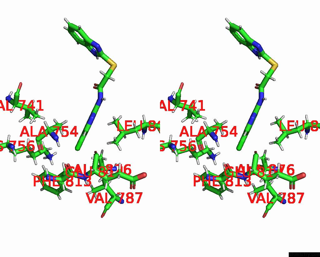
Stereo pair view
|
A full contact list of Chlorine with other atoms in the Cl binding
site number 2 of Structure of DDB1 Bound to DCEMM3-Engaged CDK12-Cyclin K within 5.0Å range:
probe
|
atom
|
residue
|
distance (Å)
|
B
|
Occ
|
E:Cl1101
b:116.2
occ:1.00
|
CL1
|
E:RKO1101
|
0.0
|
116.2
|
1.0
|
C4
|
E:RKO1101
|
1.7
|
109.6
|
1.0
|
C3
|
E:RKO1101
|
2.7
|
136.5
|
1.0
|
C5
|
E:RKO1101
|
2.7
|
97.8
|
1.0
|
H2
|
E:RKO1101
|
2.8
|
163.8
|
0.0
|
H3
|
E:RKO1101
|
2.8
|
117.4
|
0.0
|
HB2
|
E:PHE813
|
3.0
|
131.3
|
0.0
|
HB3
|
E:PHE813
|
3.2
|
131.3
|
0.0
|
HG21
|
E:VAL787
|
3.3
|
129.8
|
0.0
|
HB3
|
E:ALA754
|
3.5
|
147.9
|
0.0
|
CB
|
E:PHE813
|
3.5
|
109.4
|
1.0
|
CG
|
E:PHE813
|
3.8
|
108.5
|
1.0
|
O
|
E:GLU814
|
3.9
|
132.2
|
1.0
|
N2
|
E:RKO1101
|
4.0
|
143.8
|
1.0
|
C6
|
E:RKO1101
|
4.0
|
110.0
|
1.0
|
HD11
|
E:LEU866
|
4.0
|
141.8
|
0.0
|
CG2
|
E:VAL787
|
4.1
|
108.2
|
1.0
|
HG22
|
E:VAL787
|
4.2
|
129.8
|
0.0
|
HD1
|
E:PHE813
|
4.2
|
132.2
|
0.0
|
CD1
|
E:PHE813
|
4.2
|
110.2
|
1.0
|
HE3
|
E:LYS756
|
4.2
|
161.1
|
0.0
|
HD13
|
E:LEU866
|
4.2
|
141.8
|
0.0
|
CB
|
E:ALA754
|
4.4
|
123.2
|
1.0
|
C2
|
E:RKO1101
|
4.4
|
131.9
|
1.0
|
CD2
|
E:PHE813
|
4.4
|
124.6
|
1.0
|
HG23
|
E:VAL787
|
4.5
|
129.8
|
0.0
|
CD1
|
E:LEU866
|
4.5
|
118.2
|
1.0
|
HB1
|
E:ALA754
|
4.6
|
147.9
|
0.0
|
HD2
|
E:PHE813
|
4.6
|
149.5
|
0.0
|
HB1
|
E:ALA876
|
4.7
|
127.7
|
0.0
|
HB2
|
E:ALA754
|
4.7
|
147.9
|
0.0
|
H4
|
E:RKO1101
|
4.7
|
132.0
|
0.0
|
HG11
|
E:VAL741
|
4.8
|
130.6
|
0.0
|
HG3
|
E:LYS756
|
4.8
|
142.9
|
0.0
|
O
|
E:ALA754
|
4.9
|
102.0
|
1.0
|
HD12
|
E:LEU866
|
4.9
|
141.8
|
0.0
|
CA
|
E:PHE813
|
4.9
|
119.5
|
1.0
|
HB2
|
E:ALA876
|
5.0
|
127.7
|
0.0
|
|
Chlorine binding site 3 out
of 3 in 8buc
Go back to
Chlorine Binding Sites List in 8buc
Chlorine binding site 3 out
of 3 in the Structure of DDB1 Bound to DCEMM3-Engaged CDK12-Cyclin K
Mono view
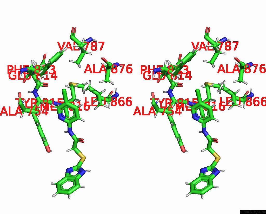
Stereo pair view
|
A full contact list of Chlorine with other atoms in the Cl binding
site number 3 of Structure of DDB1 Bound to DCEMM3-Engaged CDK12-Cyclin K within 5.0Å range:
probe
|
atom
|
residue
|
distance (Å)
|
B
|
Occ
|
H:Cl1101
b:102.1
occ:1.00
|
CL1
|
H:RKO1101
|
0.0
|
102.1
|
1.0
|
C4
|
H:RKO1101
|
1.7
|
114.5
|
1.0
|
C3
|
H:RKO1101
|
2.7
|
132.5
|
1.0
|
C5
|
H:RKO1101
|
2.7
|
109.7
|
1.0
|
H2
|
H:RKO1101
|
2.8
|
159.0
|
0.0
|
H3
|
H:RKO1101
|
2.8
|
131.6
|
0.0
|
HB3
|
H:PHE813
|
3.0
|
136.6
|
0.0
|
HD11
|
H:LEU866
|
3.4
|
129.2
|
0.0
|
HB2
|
H:PHE813
|
3.5
|
136.6
|
0.0
|
HB3
|
H:ALA754
|
3.5
|
147.1
|
0.0
|
CB
|
H:PHE813
|
3.6
|
113.9
|
1.0
|
HG21
|
H:VAL787
|
3.6
|
114.7
|
0.0
|
O
|
H:GLU814
|
3.7
|
151.5
|
1.0
|
HD13
|
H:LEU866
|
3.7
|
129.2
|
0.0
|
N2
|
H:RKO1101
|
4.0
|
133.3
|
1.0
|
C6
|
H:RKO1101
|
4.0
|
123.8
|
1.0
|
CD1
|
H:LEU866
|
4.0
|
107.7
|
1.0
|
HG22
|
H:VAL787
|
4.0
|
114.7
|
0.0
|
CG
|
H:PHE813
|
4.0
|
105.8
|
1.0
|
HD1
|
H:PHE813
|
4.1
|
135.8
|
0.0
|
CG2
|
H:VAL787
|
4.2
|
95.5
|
1.0
|
HG23
|
H:VAL787
|
4.3
|
114.7
|
0.0
|
CD1
|
H:PHE813
|
4.3
|
113.2
|
1.0
|
CB
|
H:ALA754
|
4.3
|
122.5
|
1.0
|
C2
|
H:RKO1101
|
4.4
|
130.0
|
1.0
|
HD12
|
H:LEU866
|
4.4
|
129.2
|
0.0
|
HB2
|
H:ALA754
|
4.5
|
147.1
|
0.0
|
HA
|
H:TYR815
|
4.6
|
152.8
|
0.0
|
H
|
H:MET816
|
4.6
|
146.2
|
0.0
|
HB1
|
H:ALA754
|
4.6
|
147.1
|
0.0
|
HB1
|
H:ALA876
|
4.7
|
157.6
|
0.0
|
HD21
|
H:LEU866
|
4.7
|
124.9
|
0.0
|
H4
|
H:RKO1101
|
4.7
|
148.6
|
0.0
|
C
|
H:GLU814
|
4.8
|
135.4
|
1.0
|
CD2
|
H:PHE813
|
4.8
|
110.8
|
1.0
|
CA
|
H:PHE813
|
5.0
|
111.2
|
1.0
|
|
Reference:
Z.Kozicka,
G.Kempf,
G.Petzold,
N.H.Thoma.
Structure of DDB1 Bound to Z11-Engaged CDK12-Cyclin K To Be Published.
Page generated: Tue Jul 30 07:26:43 2024
|