Chlorine »
PDB 8h8q-8huv »
8hul »
Chlorine in PDB 8hul: X-Ray Structure of Human Ppar Delta Ligand Binding Domain-Lanifibranor Co-Crystals Obtained By Co-Crystallization
Protein crystallography data
The structure of X-Ray Structure of Human Ppar Delta Ligand Binding Domain-Lanifibranor Co-Crystals Obtained By Co-Crystallization, PDB code: 8hul
was solved by
S.Kamata,
A.Honda,
Y.Machida,
K.Uchii,
Y.Shiiyama,
R.Masuda,
T.Oyama,
I.Ishii,
with X-Ray Crystallography technique. A brief refinement statistics is given in the table below:
| Resolution Low / High (Å) | 38.81 / 2.46 |
| Space group | P 1 21 1 |
| Cell size a, b, c (Å), α, β, γ (°) | 39.166, 94.948, 96.932, 90, 97.78, 90 |
| R / Rfree (%) | 21.1 / 24.3 |
Chlorine Binding Sites:
The binding sites of Chlorine atom in the X-Ray Structure of Human Ppar Delta Ligand Binding Domain-Lanifibranor Co-Crystals Obtained By Co-Crystallization
(pdb code 8hul). This binding sites where shown within
5.0 Angstroms radius around Chlorine atom.
In total 2 binding sites of Chlorine where determined in the X-Ray Structure of Human Ppar Delta Ligand Binding Domain-Lanifibranor Co-Crystals Obtained By Co-Crystallization, PDB code: 8hul:
Jump to Chlorine binding site number: 1; 2;
In total 2 binding sites of Chlorine where determined in the X-Ray Structure of Human Ppar Delta Ligand Binding Domain-Lanifibranor Co-Crystals Obtained By Co-Crystallization, PDB code: 8hul:
Jump to Chlorine binding site number: 1; 2;
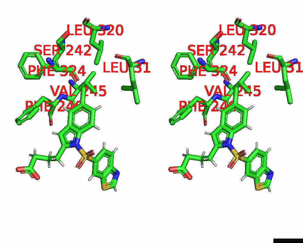
Chlorine binding site 1 out of 2 in 8hul
Go back to
Chlorine binding site 1 out
of 2 in the X-Ray Structure of Human Ppar Delta Ligand Binding Domain-Lanifibranor Co-Crystals Obtained By Co-Crystallization

Mono view

Stereo pair view
Mono view
Stereo pair view
A full contact list of Chlorine with other atoms in the Cl binding
site number 1 of X-Ray Structure of Human Ppar Delta Ligand Binding Domain-Lanifibranor Co-Crystals Obtained By Co-Crystallization within 5.0Å range:
|
Chlorine binding site 2 out of 2 in 8hul
Go back to
Chlorine binding site 2 out
of 2 in the X-Ray Structure of Human Ppar Delta Ligand Binding Domain-Lanifibranor Co-Crystals Obtained By Co-Crystallization

Mono view
Stereo pair view
Mono view
Stereo pair view
A full contact list of Chlorine with other atoms in the Cl binding
site number 2 of X-Ray Structure of Human Ppar Delta Ligand Binding Domain-Lanifibranor Co-Crystals Obtained By Co-Crystallization within 5.0Å range:
|
Reference:
S.Kamata,
A.Honda,
R.Ishikawa,
M.Akahane,
A.Fujita,
C.Kaneko,
S.Miyawaki,
Y.Habu,
Y.Shiiyama,
K.Uchii,
Y.Machida,
T.Oyama,
I.Ishii.
Functional and Structural Insights Into the Human Ppar Alpha / Delta / Gamma Targeting Preferences of Anti-Nash Investigational Drugs, Lanifibranor, Seladelpar, and Elafibranor Antioxidants V. 12 1523 2023.
ISSN: ESSN 2076-3921
DOI: 10.3390/ANTIOX12081523
Page generated: Sun Jul 13 12:07:43 2025
ISSN: ESSN 2076-3921
DOI: 10.3390/ANTIOX12081523
Last articles
F in 7MOGF in 7MOO
F in 7MML
F in 7MMI
F in 7MMK
F in 7MMJ
F in 7MMG
F in 7MMF
F in 7MMH
F in 7MMA