Chlorine »
PDB 8it6-8j20 »
8izj »
Chlorine in PDB 8izj: Crystal Structure of Escherichia Coli Adenine Phosphoribosyltransferase (Aprt) in Complex with Amp
Enzymatic activity of Crystal Structure of Escherichia Coli Adenine Phosphoribosyltransferase (Aprt) in Complex with Amp
All present enzymatic activity of Crystal Structure of Escherichia Coli Adenine Phosphoribosyltransferase (Aprt) in Complex with Amp:
2.4.2.7;
2.4.2.7;
Protein crystallography data
The structure of Crystal Structure of Escherichia Coli Adenine Phosphoribosyltransferase (Aprt) in Complex with Amp, PDB code: 8izj
was solved by
P.Yadav,
G.S.Kushwaha,
N.S.Bhavesh,
with X-Ray Crystallography technique. A brief refinement statistics is given in the table below:
| Resolution Low / High (Å) | 45.59 / 1.60 |
| Space group | P 1 21 1 |
| Cell size a, b, c (Å), α, β, γ (°) | 41.752, 86.942, 48.664, 90, 110.48, 90 |
| R / Rfree (%) | 16.5 / 19.2 |
Other elements in 8izj:
The structure of Crystal Structure of Escherichia Coli Adenine Phosphoribosyltransferase (Aprt) in Complex with Amp also contains other interesting chemical elements:
| Sodium | (Na) | 2 atoms |
Chlorine Binding Sites:
The binding sites of Chlorine atom in the Crystal Structure of Escherichia Coli Adenine Phosphoribosyltransferase (Aprt) in Complex with Amp
(pdb code 8izj). This binding sites where shown within
5.0 Angstroms radius around Chlorine atom.
In total 4 binding sites of Chlorine where determined in the Crystal Structure of Escherichia Coli Adenine Phosphoribosyltransferase (Aprt) in Complex with Amp, PDB code: 8izj:
Jump to Chlorine binding site number: 1; 2; 3; 4;
In total 4 binding sites of Chlorine where determined in the Crystal Structure of Escherichia Coli Adenine Phosphoribosyltransferase (Aprt) in Complex with Amp, PDB code: 8izj:
Jump to Chlorine binding site number: 1; 2; 3; 4;
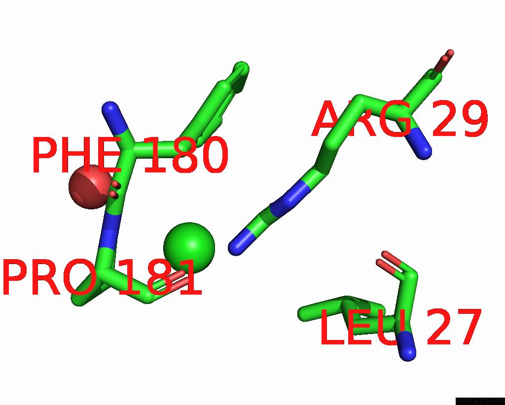
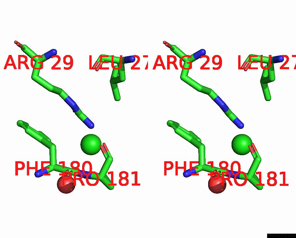
Chlorine binding site 1 out of 4 in 8izj
Go back to
Chlorine binding site 1 out
of 4 in the Crystal Structure of Escherichia Coli Adenine Phosphoribosyltransferase (Aprt) in Complex with Amp
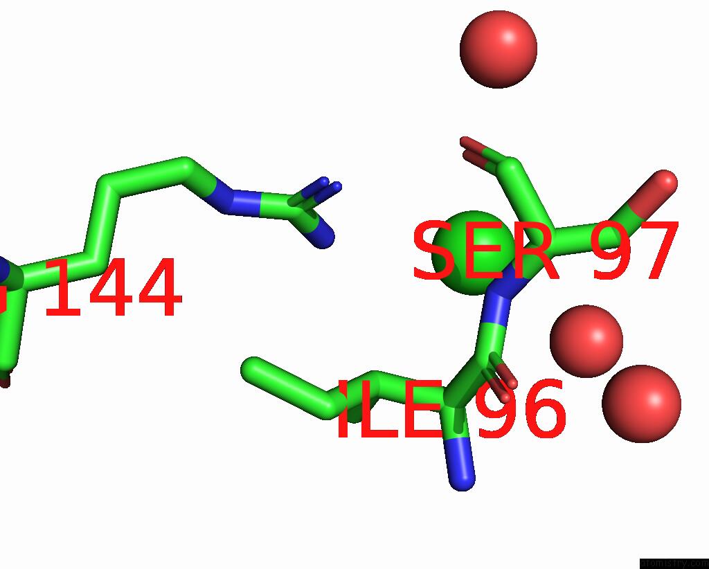
Mono view
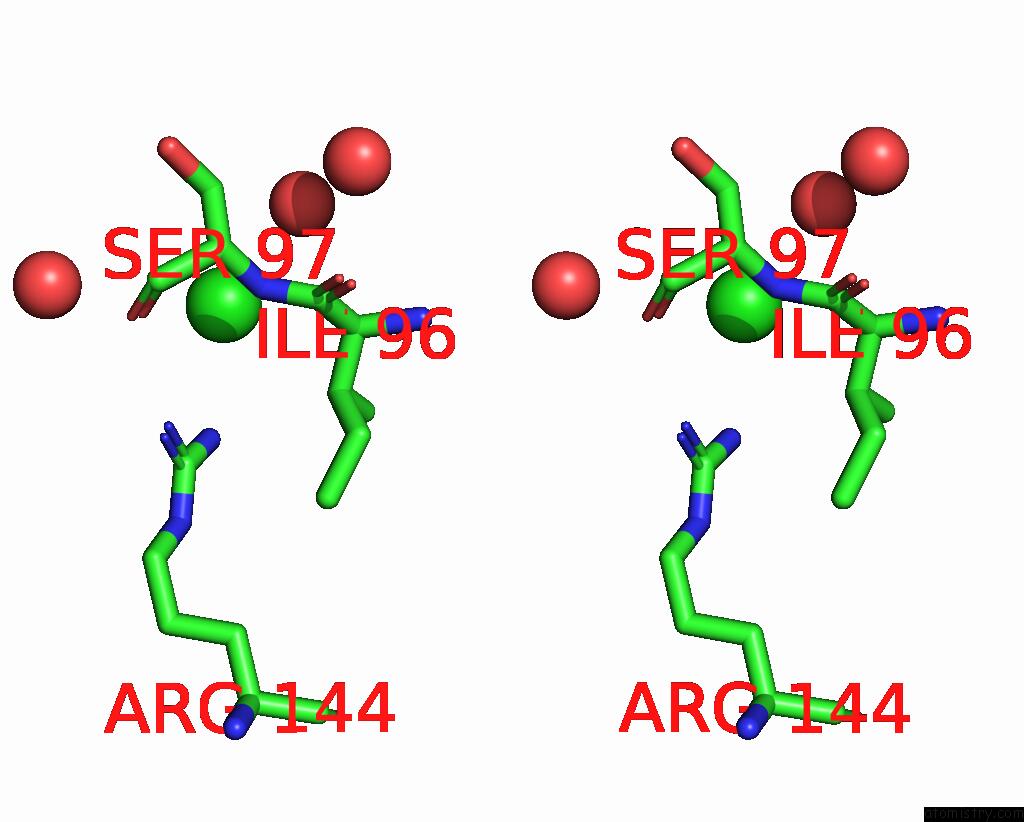
Stereo pair view
Mono view
Stereo pair view
A full contact list of Chlorine with other atoms in the Cl binding
site number 1 of Crystal Structure of Escherichia Coli Adenine Phosphoribosyltransferase (Aprt) in Complex with Amp within 5.0Å range:
|
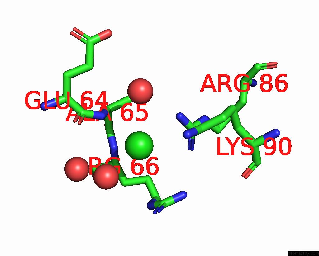
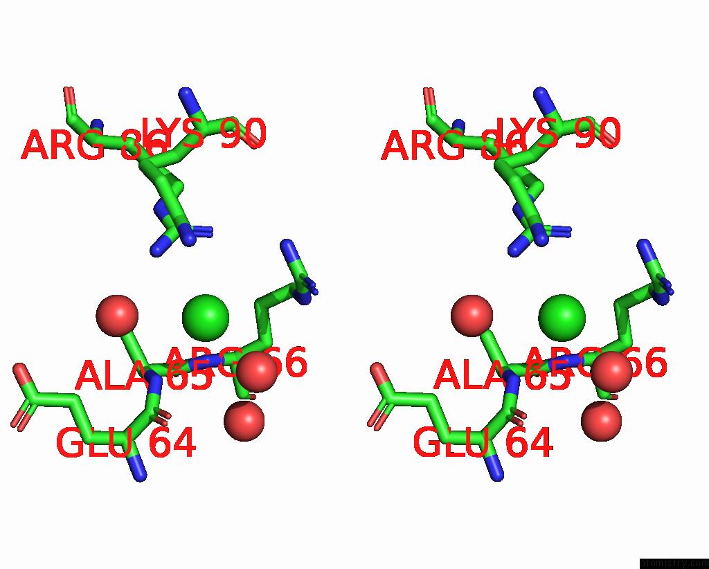
Chlorine binding site 2 out of 4 in 8izj
Go back to
Chlorine binding site 2 out
of 4 in the Crystal Structure of Escherichia Coli Adenine Phosphoribosyltransferase (Aprt) in Complex with Amp
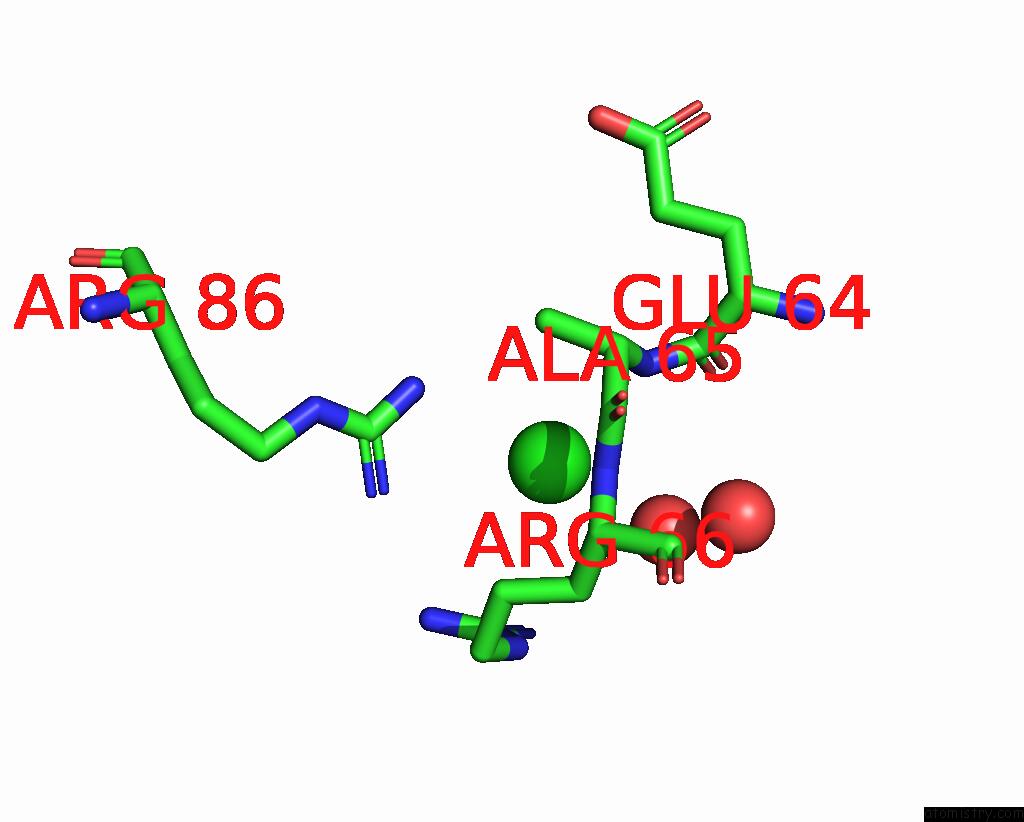
Mono view
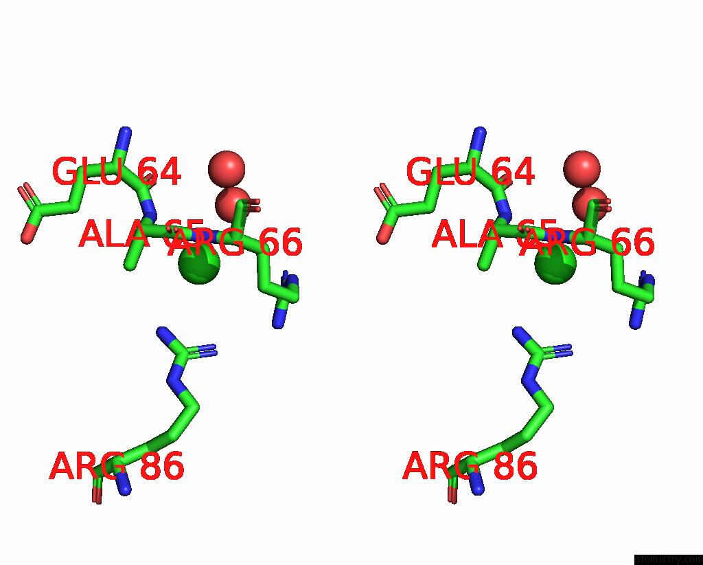
Stereo pair view
Mono view
Stereo pair view
A full contact list of Chlorine with other atoms in the Cl binding
site number 2 of Crystal Structure of Escherichia Coli Adenine Phosphoribosyltransferase (Aprt) in Complex with Amp within 5.0Å range:
|
Chlorine binding site 3 out of 4 in 8izj
Go back to
Chlorine binding site 3 out
of 4 in the Crystal Structure of Escherichia Coli Adenine Phosphoribosyltransferase (Aprt) in Complex with Amp
Mono view
Stereo pair view
Mono view
Stereo pair view
A full contact list of Chlorine with other atoms in the Cl binding
site number 3 of Crystal Structure of Escherichia Coli Adenine Phosphoribosyltransferase (Aprt) in Complex with Amp within 5.0Å range:
|
Chlorine binding site 4 out of 4 in 8izj
Go back to
Chlorine binding site 4 out
of 4 in the Crystal Structure of Escherichia Coli Adenine Phosphoribosyltransferase (Aprt) in Complex with Amp
Mono view
Stereo pair view
Mono view
Stereo pair view
A full contact list of Chlorine with other atoms in the Cl binding
site number 4 of Crystal Structure of Escherichia Coli Adenine Phosphoribosyltransferase (Aprt) in Complex with Amp within 5.0Å range:
|
Reference:
P.Yadav,
G.S.Kushwaha,
N.S.Bhavesh.
Crystal Structure of Escherichia Coli Adenine Phosphoribosyltransferase (Aprt) in Complex with Amp To Be Published.
Page generated: Sun Jul 13 12:32:18 2025
Last articles
Fe in 2J1MFe in 2J19
Fe in 2J18
Fe in 2J0P
Fe in 2IW4
Fe in 2J0D
Fe in 2IV2
Fe in 2IVP
Fe in 2IVJ
Fe in 2ISA