Chlorine »
PDB 8pow-8q2s »
8pwj »
Chlorine in PDB 8pwj: Light Structure of Sensory Rhodopsin-II Solved By Serial Millisecond Crystallography. 30-60 Milliseconds Time-Bin
Protein crystallography data
The structure of Light Structure of Sensory Rhodopsin-II Solved By Serial Millisecond Crystallography. 30-60 Milliseconds Time-Bin, PDB code: 8pwj
was solved by
R.Bosman,
G.Ortolani,
G.Branden,
R.Neutze,
with X-Ray Crystallography technique. A brief refinement statistics is given in the table below:
| Resolution Low / High (Å) | 44.88 / 2.14 |
| Space group | C 2 2 21 |
| Cell size a, b, c (Å), α, β, γ (°) | 89.75, 131.7, 51, 90, 90, 90 |
| R / Rfree (%) | 24.9 / 31.4 |
Chlorine Binding Sites:
The binding sites of Chlorine atom in the Light Structure of Sensory Rhodopsin-II Solved By Serial Millisecond Crystallography. 30-60 Milliseconds Time-Bin
(pdb code 8pwj). This binding sites where shown within
5.0 Angstroms radius around Chlorine atom.
In total only one binding site of Chlorine was determined in the Light Structure of Sensory Rhodopsin-II Solved By Serial Millisecond Crystallography. 30-60 Milliseconds Time-Bin, PDB code: 8pwj:
In total only one binding site of Chlorine was determined in the Light Structure of Sensory Rhodopsin-II Solved By Serial Millisecond Crystallography. 30-60 Milliseconds Time-Bin, PDB code: 8pwj:
Chlorine binding site 1 out of 1 in 8pwj
Go back to
Chlorine binding site 1 out
of 1 in the Light Structure of Sensory Rhodopsin-II Solved By Serial Millisecond Crystallography. 30-60 Milliseconds Time-Bin
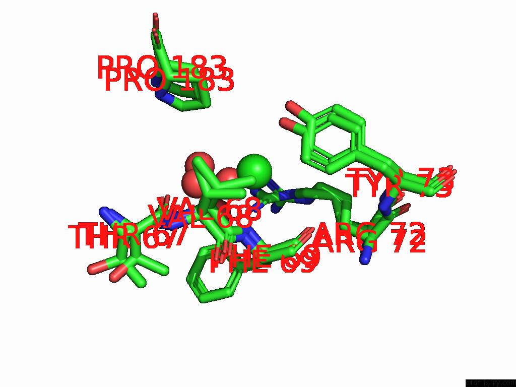
Mono view
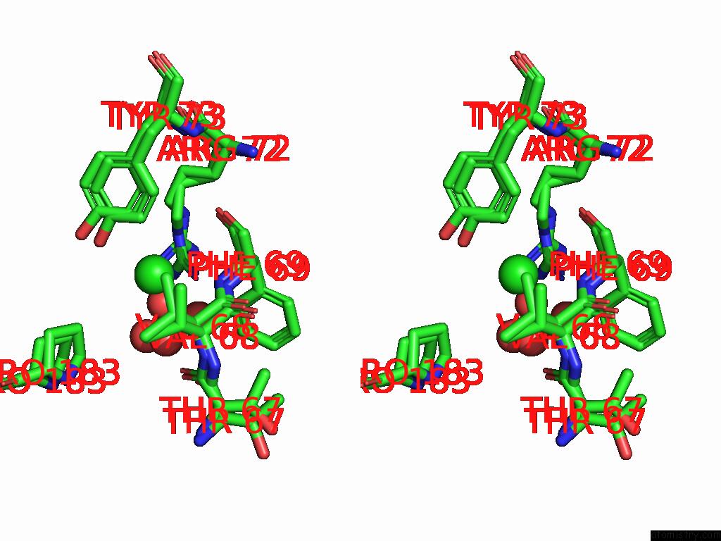
Stereo pair view
Mono view
Stereo pair view
A full contact list of Chlorine with other atoms in the Cl binding
site number 1 of Light Structure of Sensory Rhodopsin-II Solved By Serial Millisecond Crystallography. 30-60 Milliseconds Time-Bin within 5.0Å range:
|
Reference:
G.Ortolani,
R.Bosman,
G.Branden,
R.Neutze.
Structural Basis of the Prolonged Photocycle of Sensory Rhodopsin II Revealed By Serial Millisecond Crystallography To Be Published.
Page generated: Sun Feb 9 06:44:44 2025
Last articles
Cl in 2VF3Cl in 2VFV
Cl in 2VFT
Cl in 2VFS
Cl in 2VFR
Cl in 2VDE
Cl in 2VEL
Cl in 2VEC
Cl in 2VD3
Cl in 2VD6