Chlorine »
PDB 8qk4-8qsf »
8qoc »
Chlorine in PDB 8qoc: Crystal Structure of Staphylococcus Aureus Plp Synthase (PDX1)
Protein crystallography data
The structure of Crystal Structure of Staphylococcus Aureus Plp Synthase (PDX1), PDB code: 8qoc
was solved by
N.Ullah,
C.Wrenger,
C.Betzel,
with X-Ray Crystallography technique. A brief refinement statistics is given in the table below:
| Resolution Low / High (Å) | 49.80 / 2.83 |
| Space group | H 3 2 1 |
| Cell size a, b, c (Å), α, β, γ (°) | 192.419, 192.419, 448.198, 90, 90, 120 |
| R / Rfree (%) | 20.2 / 26 |
Other elements in 8qoc:
The structure of Crystal Structure of Staphylococcus Aureus Plp Synthase (PDX1) also contains other interesting chemical elements:
| Magnesium | (Mg) | 3 atoms |
Chlorine Binding Sites:
The binding sites of Chlorine atom in the Crystal Structure of Staphylococcus Aureus Plp Synthase (PDX1)
(pdb code 8qoc). This binding sites where shown within
5.0 Angstroms radius around Chlorine atom.
In total 8 binding sites of Chlorine where determined in the Crystal Structure of Staphylococcus Aureus Plp Synthase (PDX1), PDB code: 8qoc:
Jump to Chlorine binding site number: 1; 2; 3; 4; 5; 6; 7; 8;
In total 8 binding sites of Chlorine where determined in the Crystal Structure of Staphylococcus Aureus Plp Synthase (PDX1), PDB code: 8qoc:
Jump to Chlorine binding site number: 1; 2; 3; 4; 5; 6; 7; 8;
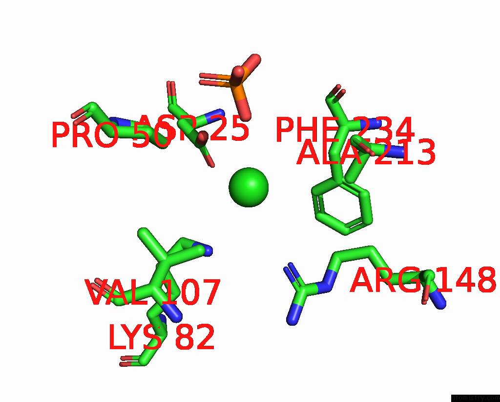
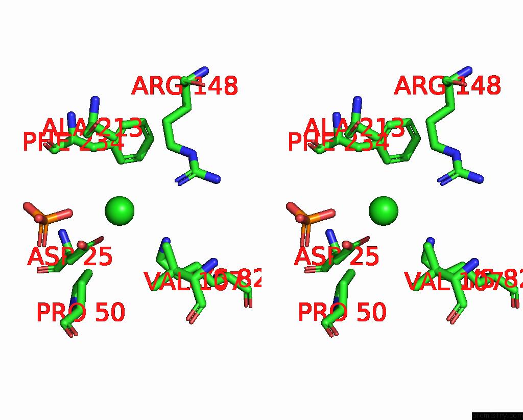
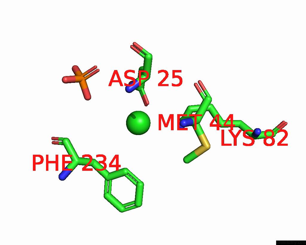
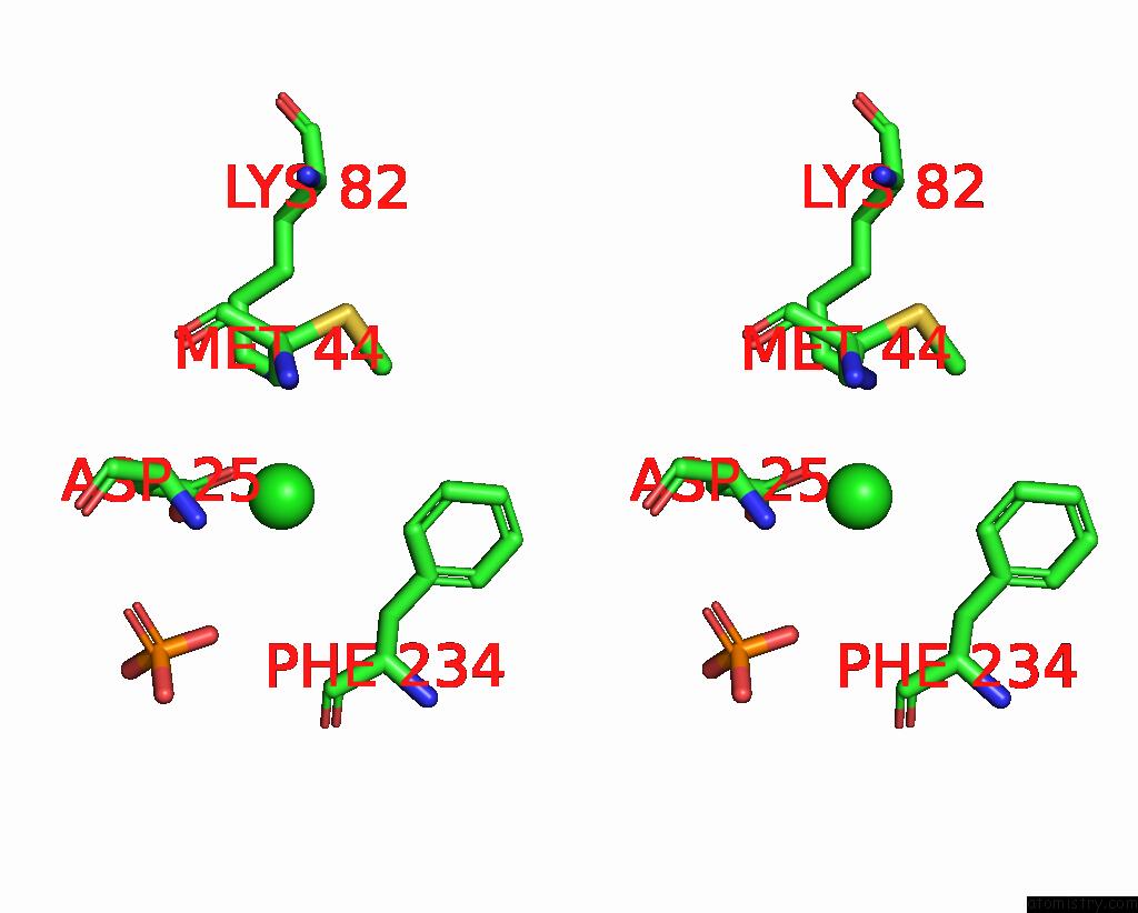
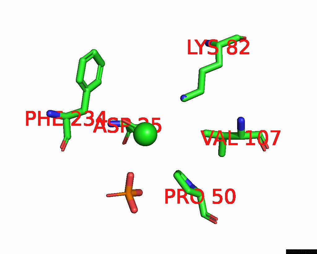
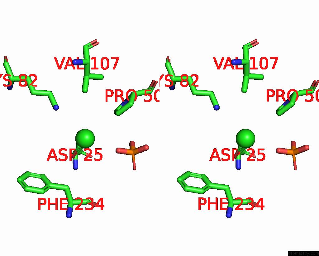
Chlorine binding site 1 out of 8 in 8qoc
Go back to
Chlorine binding site 1 out
of 8 in the Crystal Structure of Staphylococcus Aureus Plp Synthase (PDX1)
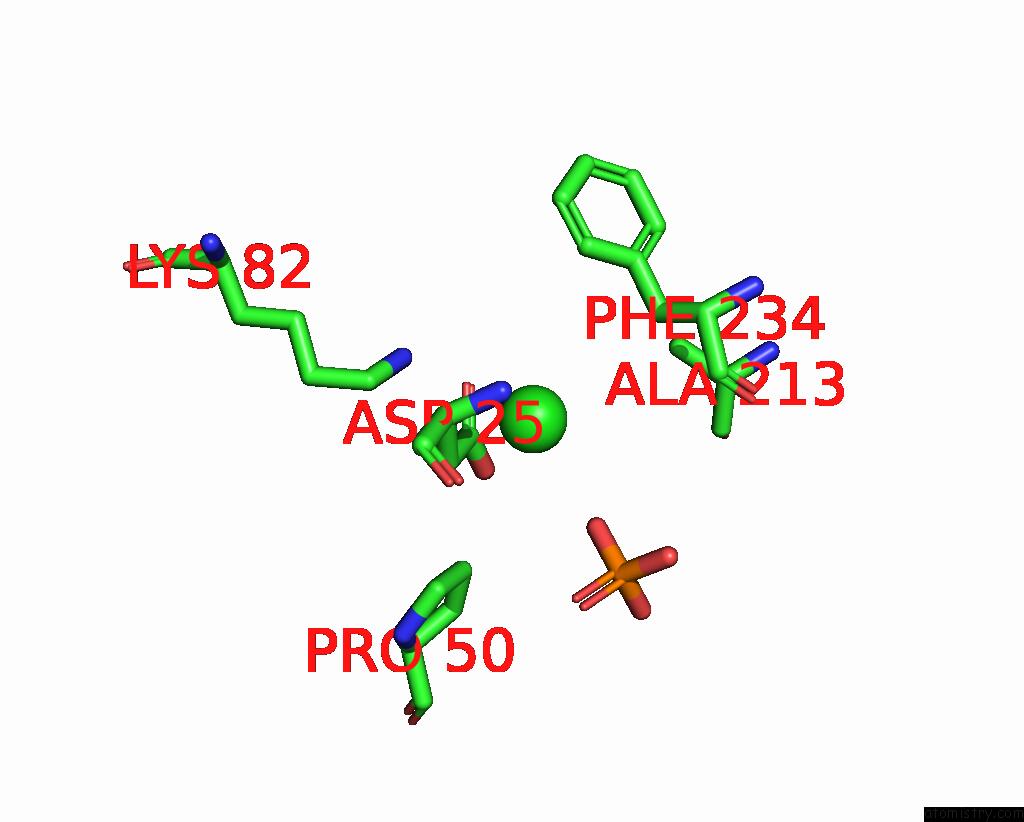
Mono view
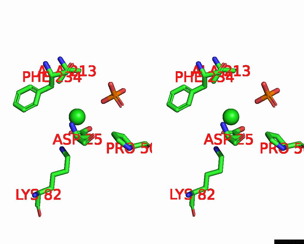
Stereo pair view
Mono view
Stereo pair view
A full contact list of Chlorine with other atoms in the Cl binding
site number 1 of Crystal Structure of Staphylococcus Aureus Plp Synthase (PDX1) within 5.0Å range:
|
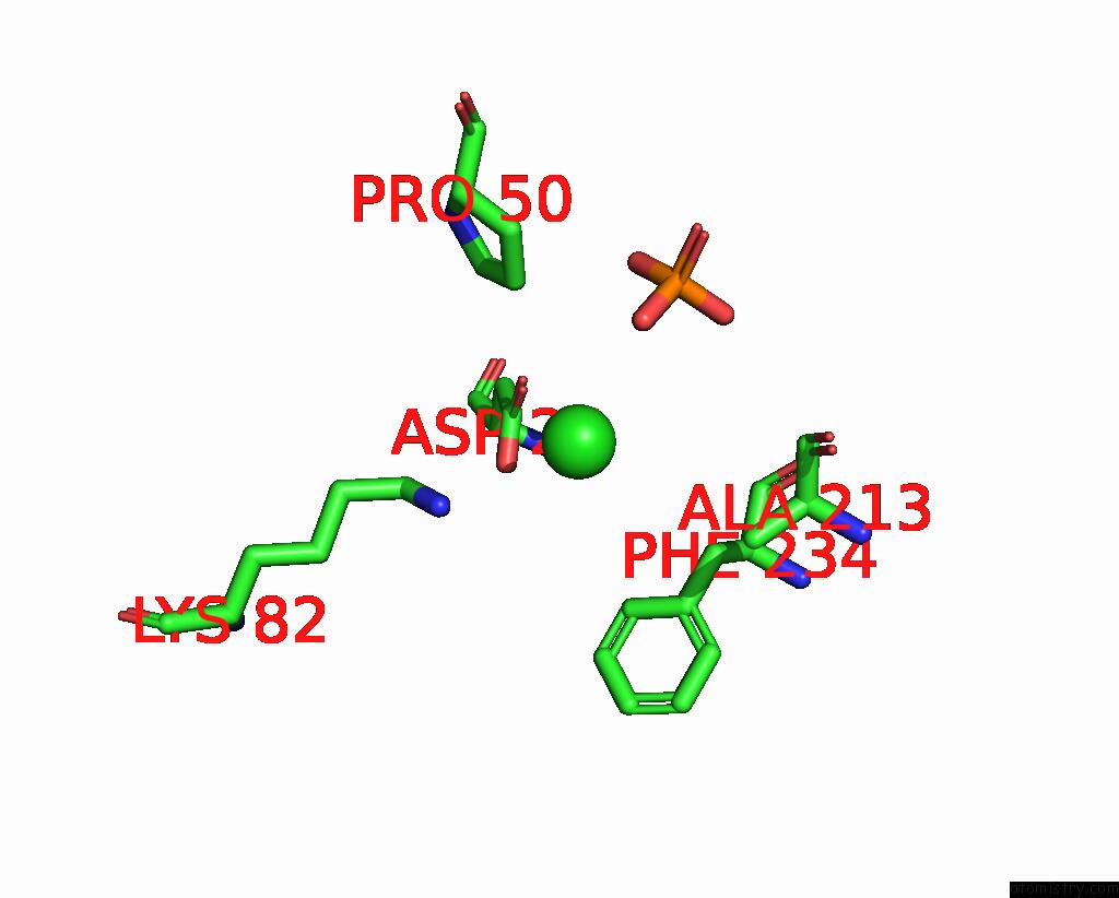
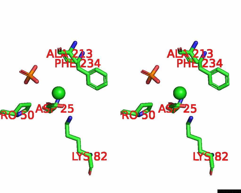
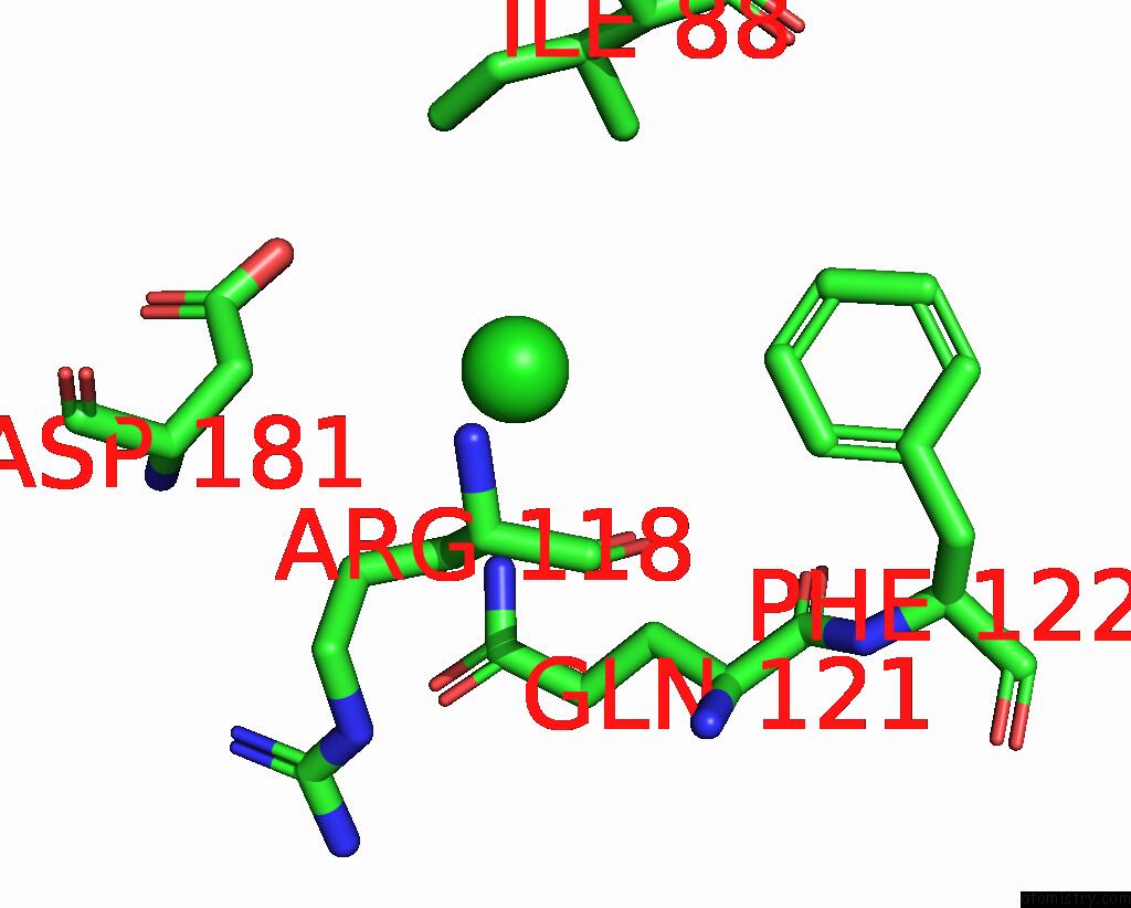
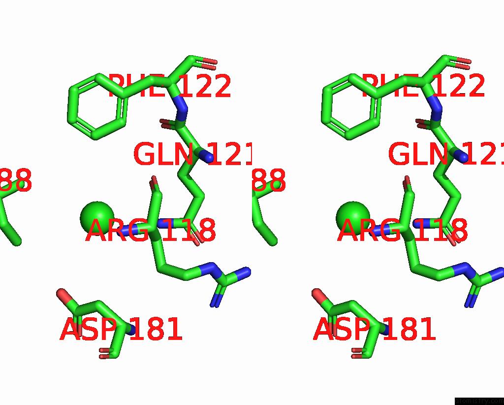
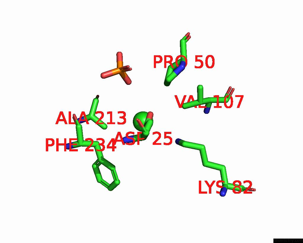
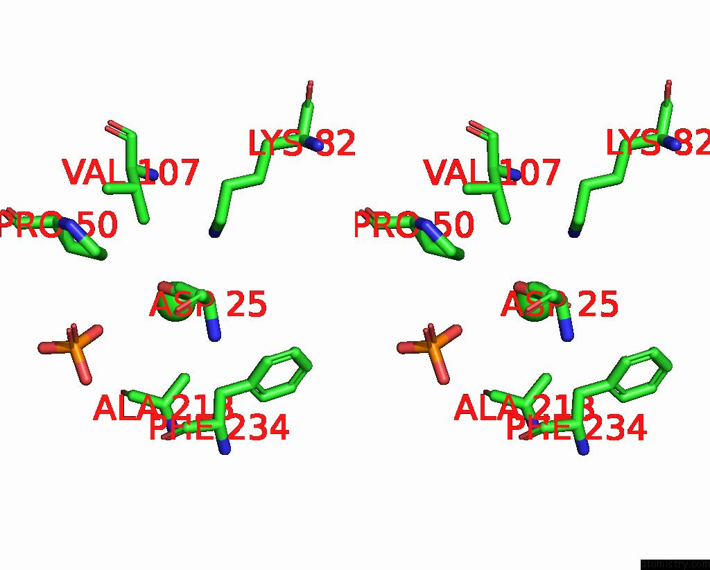
Chlorine binding site 2 out of 8 in 8qoc
Go back to
Chlorine binding site 2 out
of 8 in the Crystal Structure of Staphylococcus Aureus Plp Synthase (PDX1)
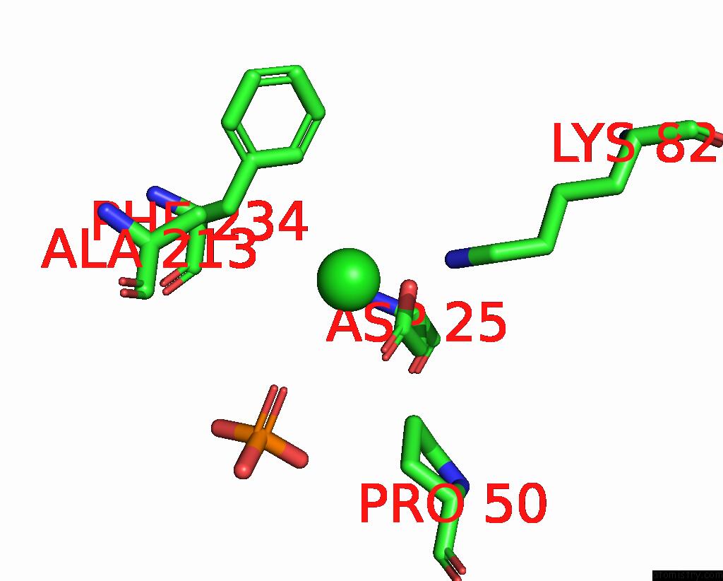
Mono view
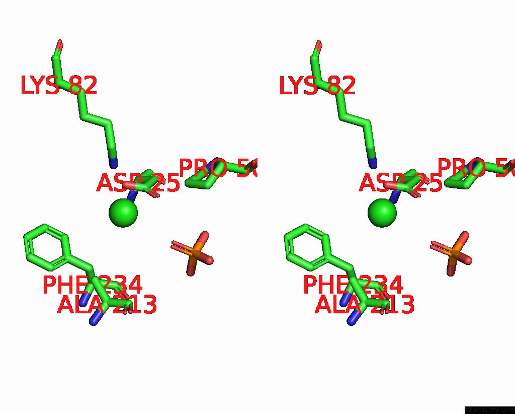
Stereo pair view
Mono view
Stereo pair view
A full contact list of Chlorine with other atoms in the Cl binding
site number 2 of Crystal Structure of Staphylococcus Aureus Plp Synthase (PDX1) within 5.0Å range:
|
Chlorine binding site 3 out of 8 in 8qoc
Go back to
Chlorine binding site 3 out
of 8 in the Crystal Structure of Staphylococcus Aureus Plp Synthase (PDX1)
Mono view
Stereo pair view
Mono view
Stereo pair view
A full contact list of Chlorine with other atoms in the Cl binding
site number 3 of Crystal Structure of Staphylococcus Aureus Plp Synthase (PDX1) within 5.0Å range:
|
Chlorine binding site 4 out of 8 in 8qoc
Go back to
Chlorine binding site 4 out
of 8 in the Crystal Structure of Staphylococcus Aureus Plp Synthase (PDX1)
Mono view
Stereo pair view
Mono view
Stereo pair view
A full contact list of Chlorine with other atoms in the Cl binding
site number 4 of Crystal Structure of Staphylococcus Aureus Plp Synthase (PDX1) within 5.0Å range:
|
Chlorine binding site 5 out of 8 in 8qoc
Go back to
Chlorine binding site 5 out
of 8 in the Crystal Structure of Staphylococcus Aureus Plp Synthase (PDX1)
Mono view
Stereo pair view
Mono view
Stereo pair view
A full contact list of Chlorine with other atoms in the Cl binding
site number 5 of Crystal Structure of Staphylococcus Aureus Plp Synthase (PDX1) within 5.0Å range:
|
Chlorine binding site 6 out of 8 in 8qoc
Go back to
Chlorine binding site 6 out
of 8 in the Crystal Structure of Staphylococcus Aureus Plp Synthase (PDX1)
Mono view
Stereo pair view
Mono view
Stereo pair view
A full contact list of Chlorine with other atoms in the Cl binding
site number 6 of Crystal Structure of Staphylococcus Aureus Plp Synthase (PDX1) within 5.0Å range:
|
Chlorine binding site 7 out of 8 in 8qoc
Go back to
Chlorine binding site 7 out
of 8 in the Crystal Structure of Staphylococcus Aureus Plp Synthase (PDX1)
Mono view
Stereo pair view
Mono view
Stereo pair view
A full contact list of Chlorine with other atoms in the Cl binding
site number 7 of Crystal Structure of Staphylococcus Aureus Plp Synthase (PDX1) within 5.0Å range:
|
Chlorine binding site 8 out of 8 in 8qoc
Go back to
Chlorine binding site 8 out
of 8 in the Crystal Structure of Staphylococcus Aureus Plp Synthase (PDX1)
Mono view
Stereo pair view
Mono view
Stereo pair view
A full contact list of Chlorine with other atoms in the Cl binding
site number 8 of Crystal Structure of Staphylococcus Aureus Plp Synthase (PDX1) within 5.0Å range:
|
Reference:
A.L.C.Barra,
N.Ullah,
H.Brognaro,
R.F.Gutierrez,
C.Wrenger,
C.Betzel,
A.S.Nascimento.
Structure and Dynamics of the Staphylococcal Pyridoxal 5-Phosphate Synthase Complex Reveal Transient Interactions at the Enzyme Interface. J.Biol.Chem. V. 300 07404 2024.
ISSN: ESSN 1083-351X
PubMed: 38782204
DOI: 10.1016/J.JBC.2024.107404
Page generated: Sun Jul 13 13:37:30 2025
ISSN: ESSN 1083-351X
PubMed: 38782204
DOI: 10.1016/J.JBC.2024.107404
Last articles
F in 4GG5F in 4GE6
F in 4GE5
F in 4GCA
F in 4GE2
F in 4G9C
F in 4GAB
F in 4GA8
F in 4G90
F in 4G8R