Chlorine »
PDB 8ri3-8rus »
8rs5 »
Chlorine in PDB 8rs5: Crystal Structure of Methanopyrus Kandleri Malate Dehydrogenase Mutant 4
Enzymatic activity of Crystal Structure of Methanopyrus Kandleri Malate Dehydrogenase Mutant 4
All present enzymatic activity of Crystal Structure of Methanopyrus Kandleri Malate Dehydrogenase Mutant 4:
1.1.1.299;
1.1.1.299;
Protein crystallography data
The structure of Crystal Structure of Methanopyrus Kandleri Malate Dehydrogenase Mutant 4, PDB code: 8rs5
was solved by
S.Coquille,
S.Engilberge,
D.Madern,
with X-Ray Crystallography technique. A brief refinement statistics is given in the table below:
| Resolution Low / High (Å) | 19.92 / 1.95 |
| Space group | P 43 21 2 |
| Cell size a, b, c (Å), α, β, γ (°) | 74.36, 74.36, 267.67, 90, 90, 90 |
| R / Rfree (%) | 25.8 / 28.3 |
Other elements in 8rs5:
The structure of Crystal Structure of Methanopyrus Kandleri Malate Dehydrogenase Mutant 4 also contains other interesting chemical elements:
| Potassium | (K) | 2 atoms |
Chlorine Binding Sites:
The binding sites of Chlorine atom in the Crystal Structure of Methanopyrus Kandleri Malate Dehydrogenase Mutant 4
(pdb code 8rs5). This binding sites where shown within
5.0 Angstroms radius around Chlorine atom.
In total 3 binding sites of Chlorine where determined in the Crystal Structure of Methanopyrus Kandleri Malate Dehydrogenase Mutant 4, PDB code: 8rs5:
Jump to Chlorine binding site number: 1; 2; 3;
In total 3 binding sites of Chlorine where determined in the Crystal Structure of Methanopyrus Kandleri Malate Dehydrogenase Mutant 4, PDB code: 8rs5:
Jump to Chlorine binding site number: 1; 2; 3;
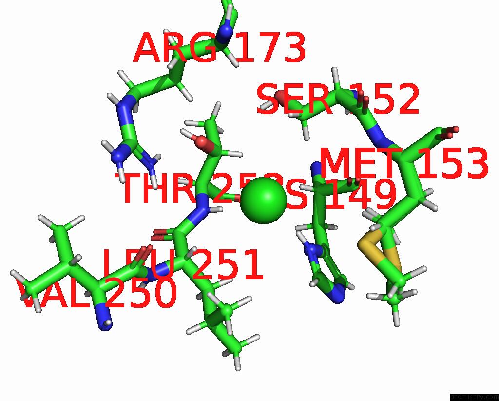
Chlorine binding site 1 out of 3 in 8rs5
Go back to
Chlorine binding site 1 out
of 3 in the Crystal Structure of Methanopyrus Kandleri Malate Dehydrogenase Mutant 4
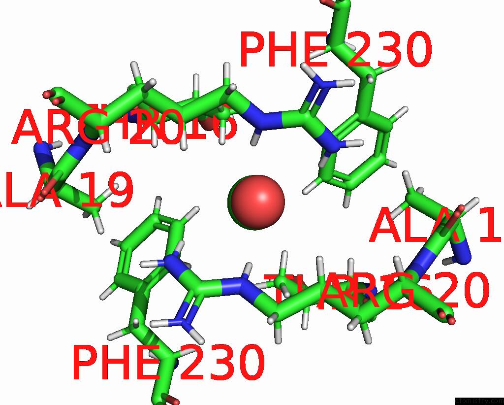
Mono view
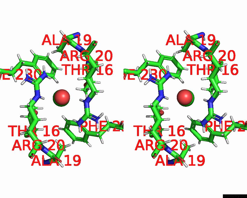
Stereo pair view
Mono view
Stereo pair view
A full contact list of Chlorine with other atoms in the Cl binding
site number 1 of Crystal Structure of Methanopyrus Kandleri Malate Dehydrogenase Mutant 4 within 5.0Å range:
|
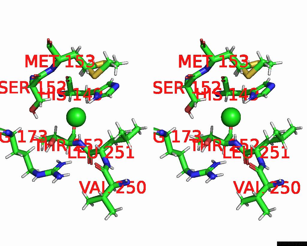
Chlorine binding site 2 out of 3 in 8rs5
Go back to
Chlorine binding site 2 out
of 3 in the Crystal Structure of Methanopyrus Kandleri Malate Dehydrogenase Mutant 4
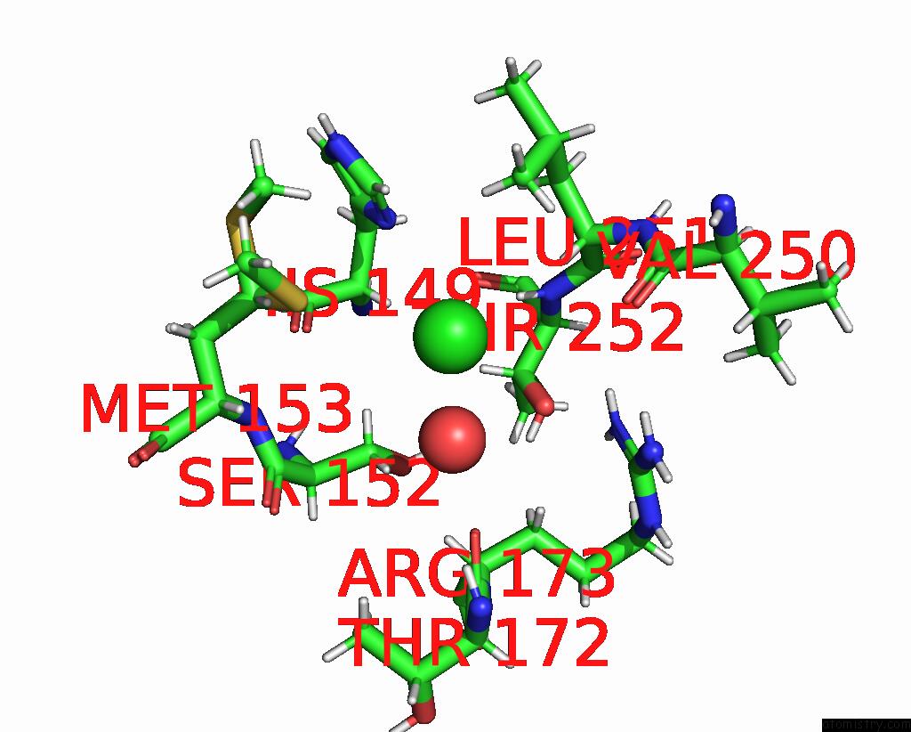
Mono view
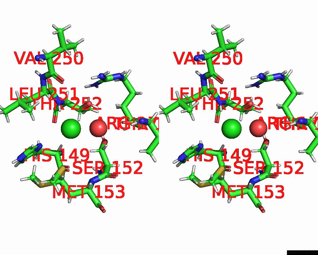
Stereo pair view
Mono view
Stereo pair view
A full contact list of Chlorine with other atoms in the Cl binding
site number 2 of Crystal Structure of Methanopyrus Kandleri Malate Dehydrogenase Mutant 4 within 5.0Å range:
|
Chlorine binding site 3 out of 3 in 8rs5
Go back to
Chlorine binding site 3 out
of 3 in the Crystal Structure of Methanopyrus Kandleri Malate Dehydrogenase Mutant 4
Mono view
Stereo pair view
Mono view
Stereo pair view
A full contact list of Chlorine with other atoms in the Cl binding
site number 3 of Crystal Structure of Methanopyrus Kandleri Malate Dehydrogenase Mutant 4 within 5.0Å range:
|
Reference:
S.Coquille,
C.Simoes Pereira,
C.Brochier-Armanet,
J.Roche,
G.Santoni,
N.Coquelle,
E.Girard,
F.Sterpone,
D.Madern.
Navigating the Conformational Landscape of An Enzyme. Stabilization of A Low Populated Conformer By Evolutionary Mutations Triggers Allostery Into A Non-Allosteric Enzyme. To Be Published.
Page generated: Sun Jul 13 14:00:04 2025
Last articles
Fe in 2YXOFe in 2YRS
Fe in 2YXC
Fe in 2YNM
Fe in 2YVJ
Fe in 2YP1
Fe in 2YU2
Fe in 2YU1
Fe in 2YQB
Fe in 2YOO