Chlorine »
PDB 8vu4-8w8v »
8w03 »
Chlorine in PDB 8w03: Crystal Structure of the Er-Alpha Ligand-Binding Domain (L372S, L536S) in Complex with K-1154
Protein crystallography data
The structure of Crystal Structure of the Er-Alpha Ligand-Binding Domain (L372S, L536S) in Complex with K-1154, PDB code: 8w03
was solved by
C.K.Min,
J.C.Nwachukwu,
Y.Hou,
R.J.Russo,
A.Papa,
J.Min,
R.Peng,
S.H.Kim,
Y.Ziegler,
E.S.Rangarajan,
T.Izard,
B.S.Katzenellenbogen,
J.A.Katzenellenbogen,
K.W.Nettles,
with X-Ray Crystallography technique. A brief refinement statistics is given in the table below:
| Resolution Low / High (Å) | 38.36 / 1.68 |
| Space group | P 1 |
| Cell size a, b, c (Å), α, β, γ (°) | 53.489, 58.771, 93.208, 86.61, 75.17, 62.96 |
| R / Rfree (%) | 20.1 / 24.5 |
Other elements in 8w03:
The structure of Crystal Structure of the Er-Alpha Ligand-Binding Domain (L372S, L536S) in Complex with K-1154 also contains other interesting chemical elements:
| Fluorine | (F) | 3 atoms |
Chlorine Binding Sites:
The binding sites of Chlorine atom in the Crystal Structure of the Er-Alpha Ligand-Binding Domain (L372S, L536S) in Complex with K-1154
(pdb code 8w03). This binding sites where shown within
5.0 Angstroms radius around Chlorine atom.
In total 3 binding sites of Chlorine where determined in the Crystal Structure of the Er-Alpha Ligand-Binding Domain (L372S, L536S) in Complex with K-1154, PDB code: 8w03:
Jump to Chlorine binding site number: 1; 2; 3;
In total 3 binding sites of Chlorine where determined in the Crystal Structure of the Er-Alpha Ligand-Binding Domain (L372S, L536S) in Complex with K-1154, PDB code: 8w03:
Jump to Chlorine binding site number: 1; 2; 3;
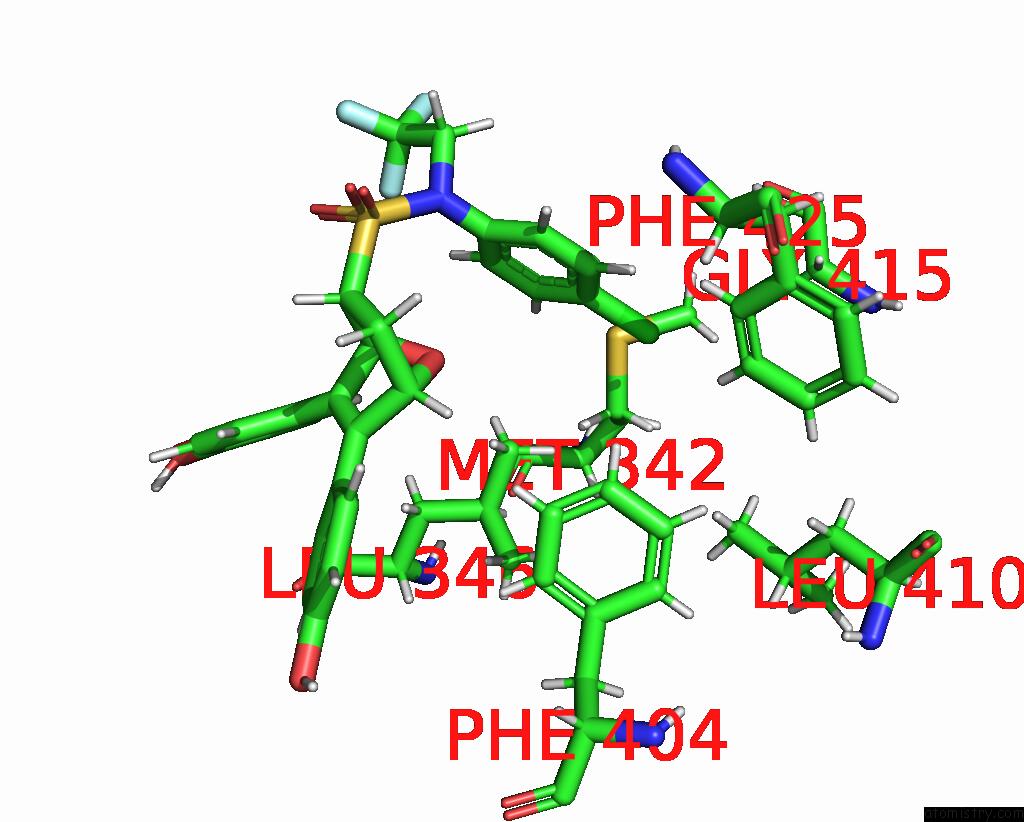
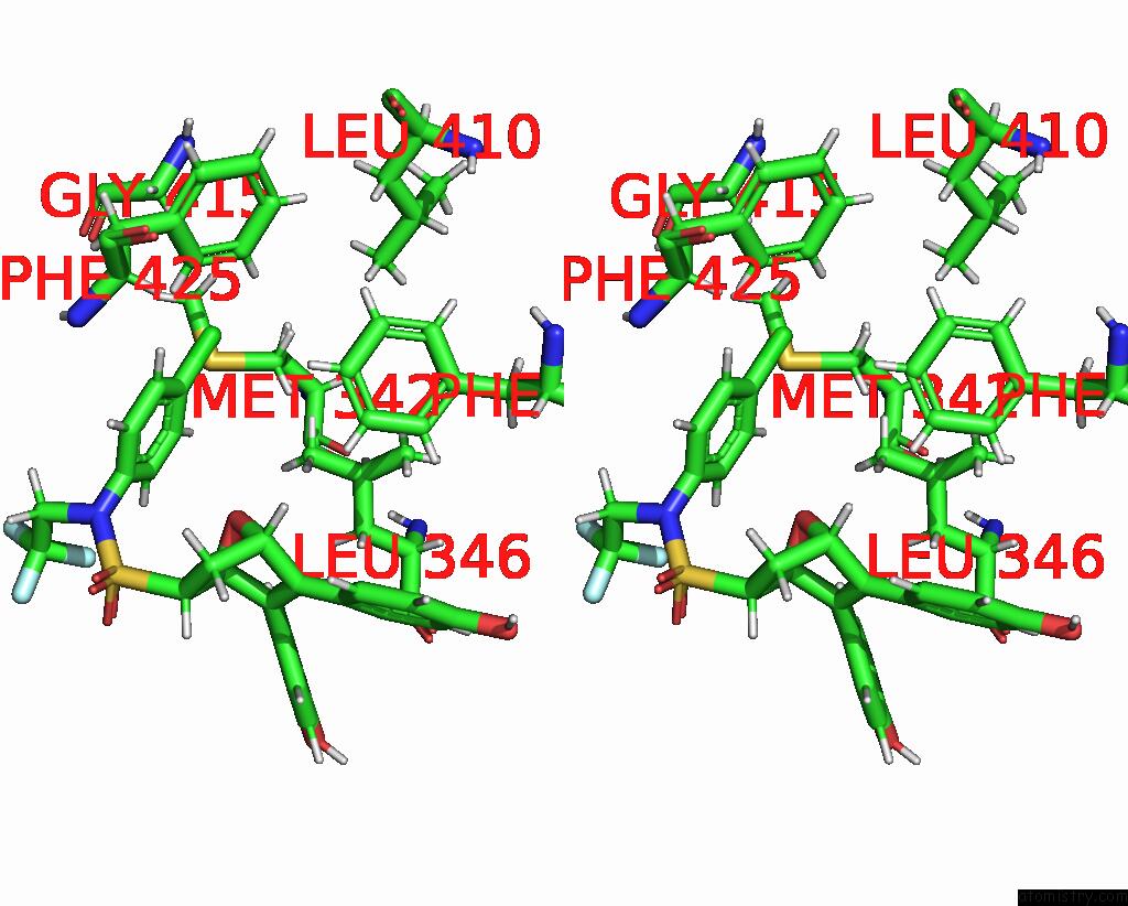
Chlorine binding site 1 out of 3 in 8w03
Go back to
Chlorine binding site 1 out
of 3 in the Crystal Structure of the Er-Alpha Ligand-Binding Domain (L372S, L536S) in Complex with K-1154
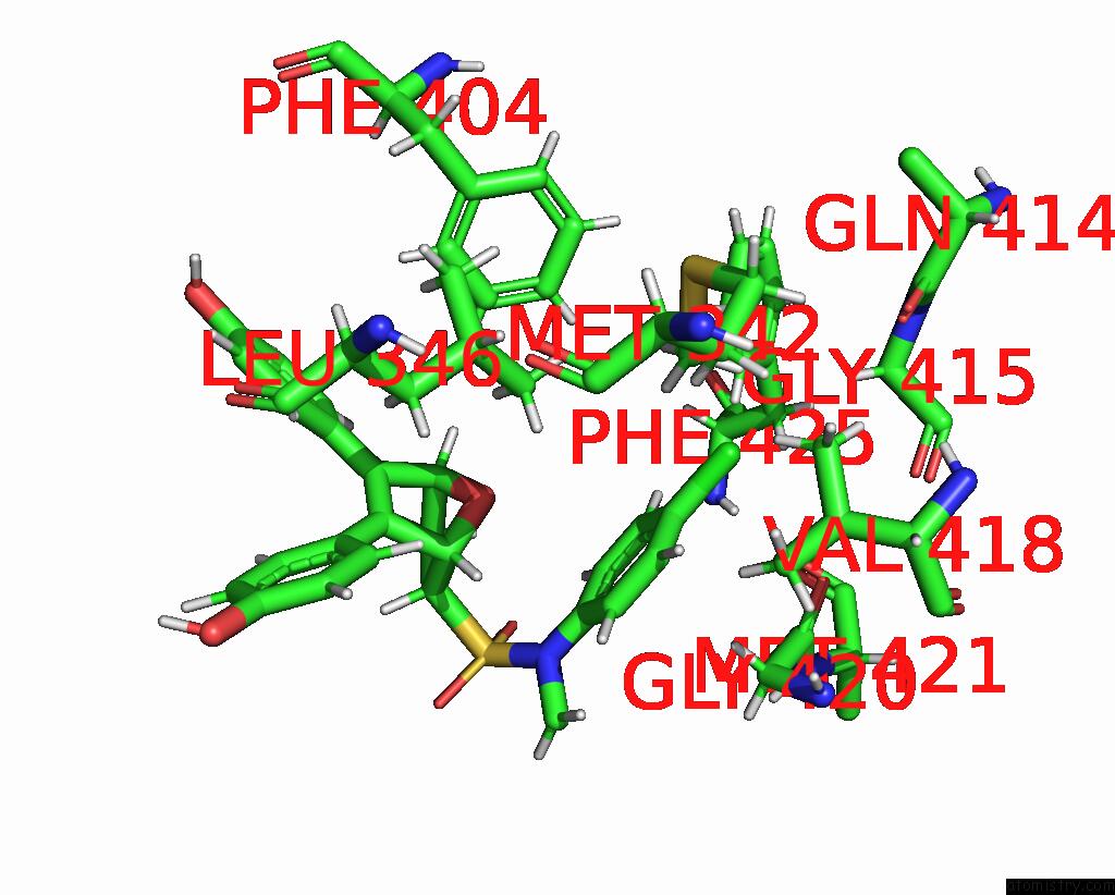
Mono view
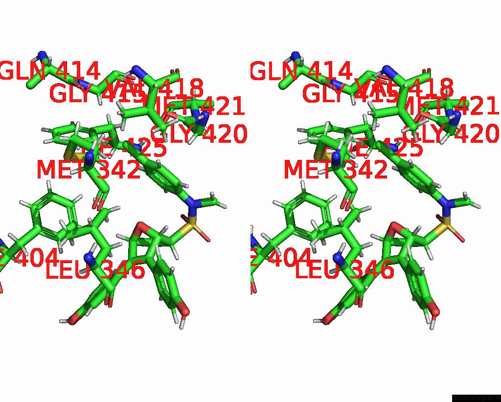
Stereo pair view
Mono view
Stereo pair view
A full contact list of Chlorine with other atoms in the Cl binding
site number 1 of Crystal Structure of the Er-Alpha Ligand-Binding Domain (L372S, L536S) in Complex with K-1154 within 5.0Å range:
|
Chlorine binding site 2 out of 3 in 8w03
Go back to
Chlorine binding site 2 out
of 3 in the Crystal Structure of the Er-Alpha Ligand-Binding Domain (L372S, L536S) in Complex with K-1154
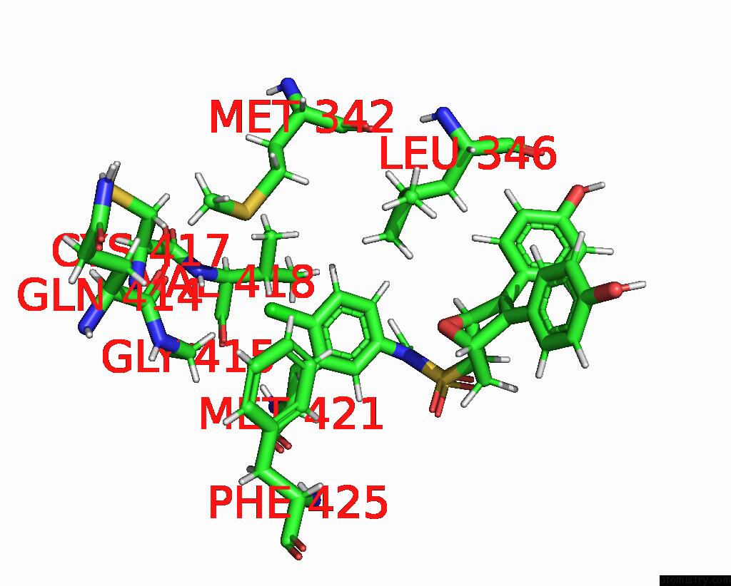
Mono view
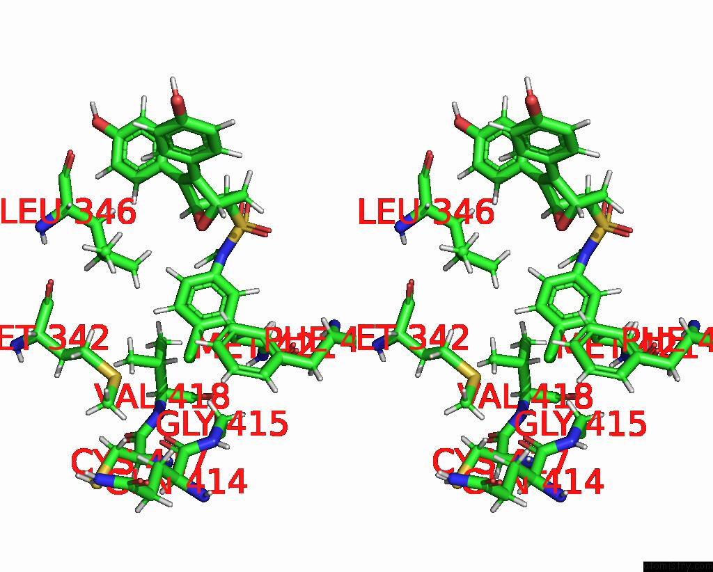
Stereo pair view
Mono view
Stereo pair view
A full contact list of Chlorine with other atoms in the Cl binding
site number 2 of Crystal Structure of the Er-Alpha Ligand-Binding Domain (L372S, L536S) in Complex with K-1154 within 5.0Å range:
|
Chlorine binding site 3 out of 3 in 8w03
Go back to
Chlorine binding site 3 out
of 3 in the Crystal Structure of the Er-Alpha Ligand-Binding Domain (L372S, L536S) in Complex with K-1154
Mono view
Stereo pair view
Mono view
Stereo pair view
A full contact list of Chlorine with other atoms in the Cl binding
site number 3 of Crystal Structure of the Er-Alpha Ligand-Binding Domain (L372S, L536S) in Complex with K-1154 within 5.0Å range:
|
Reference:
C.K.Min,
J.C.Nwachukwu,
Y.Hou,
R.J.Russo,
A.Papa,
J.Min,
R.Peng,
S.H.Kim,
Y.Ziegler,
E.S.Rangarajan,
T.Izard,
B.S.Katzenellenbogen,
J.A.Katzenellenbogen,
K.W.Nettles.
Asymmetric Allostery in Estrogen Receptor-Alpha Homodimers Drives Responses to the Ensemble of Estrogens in the Hormonal Milieu. Proc.Natl.Acad.Sci.Usa V. 121 44121 2024.
ISSN: ESSN 1091-6490
PubMed: 38830107
DOI: 10.1073/PNAS.2321344121
Page generated: Sun Jul 13 15:26:31 2025
ISSN: ESSN 1091-6490
PubMed: 38830107
DOI: 10.1073/PNAS.2321344121
Last articles
Fe in 2YXOFe in 2YRS
Fe in 2YXC
Fe in 2YNM
Fe in 2YVJ
Fe in 2YP1
Fe in 2YU2
Fe in 2YU1
Fe in 2YQB
Fe in 2YOO