Chlorine »
PDB 8wtq-8xar »
8x7x »
Chlorine in PDB 8x7x: Crystal Structure of Sads-Cov Fusion Core
Protein crystallography data
The structure of Crystal Structure of Sads-Cov Fusion Core, PDB code: 8x7x
was solved by
L.Yan,
with X-Ray Crystallography technique. A brief refinement statistics is given in the table below:
| Resolution Low / High (Å) | 28.56 / 2.59 |
| Space group | H 3 2 |
| Cell size a, b, c (Å), α, β, γ (°) | 45.176, 45.176, 418.012, 90, 90, 120 |
| R / Rfree (%) | 23.5 / 28.4 |
Chlorine Binding Sites:
The binding sites of Chlorine atom in the Crystal Structure of Sads-Cov Fusion Core
(pdb code 8x7x). This binding sites where shown within
5.0 Angstroms radius around Chlorine atom.
In total only one binding site of Chlorine was determined in the Crystal Structure of Sads-Cov Fusion Core, PDB code: 8x7x:
In total only one binding site of Chlorine was determined in the Crystal Structure of Sads-Cov Fusion Core, PDB code: 8x7x:
Chlorine binding site 1 out of 1 in 8x7x
Go back to
Chlorine binding site 1 out
of 1 in the Crystal Structure of Sads-Cov Fusion Core
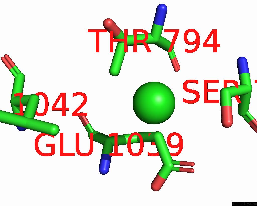
Mono view
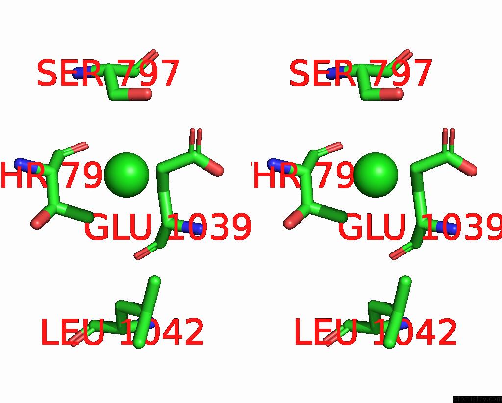
Stereo pair view
Mono view
Stereo pair view
A full contact list of Chlorine with other atoms in the Cl binding
site number 1 of Crystal Structure of Sads-Cov Fusion Core within 5.0Å range:
|
Reference:
F.Wang,
G.Yang,
L.Yan.
Crystal Structures of Fusion Cores From Ccov-Hupn-2018 and Sads-Cov Viruses V. 16 2024.
ISSN: ESSN 1999-4915
DOI: 10.3390/V16020272
Page generated: Sun Jul 13 15:40:07 2025
ISSN: ESSN 1999-4915
DOI: 10.3390/V16020272
Last articles
F in 4F9MF in 4F60
F in 4F5Q
F in 4F5O
F in 4F5N
F in 4F4Q
F in 4F4P
F in 4F2A
F in 4F2Y
F in 4E99