Chlorine »
PDB 8xb0-8yil »
8xf9 »
Chlorine in PDB 8xf9: High-Resolution Structure of the Siderophore Periplasmic Binding Protein Ftsb Mutant Y137A From Streptococcus Pyogenes
Protein crystallography data
The structure of High-Resolution Structure of the Siderophore Periplasmic Binding Protein Ftsb Mutant Y137A From Streptococcus Pyogenes, PDB code: 8xf9
was solved by
J.M.M.Caaveiro,
J.Fernandez-Perez,
K.Tsumoto,
with X-Ray Crystallography technique. A brief refinement statistics is given in the table below:
| Resolution Low / High (Å) | 40.40 / 1.15 |
| Space group | P 65 |
| Cell size a, b, c (Å), α, β, γ (°) | 75.847, 75.847, 102.622, 90, 90, 120 |
| R / Rfree (%) | 13.1 / 14.7 |
Other elements in 8xf9:
The structure of High-Resolution Structure of the Siderophore Periplasmic Binding Protein Ftsb Mutant Y137A From Streptococcus Pyogenes also contains other interesting chemical elements:
| Sodium | (Na) | 2 atoms |
| Zinc | (Zn) | 7 atoms |
Chlorine Binding Sites:
The binding sites of Chlorine atom in the High-Resolution Structure of the Siderophore Periplasmic Binding Protein Ftsb Mutant Y137A From Streptococcus Pyogenes
(pdb code 8xf9). This binding sites where shown within
5.0 Angstroms radius around Chlorine atom.
In total only one binding site of Chlorine was determined in the High-Resolution Structure of the Siderophore Periplasmic Binding Protein Ftsb Mutant Y137A From Streptococcus Pyogenes, PDB code: 8xf9:
In total only one binding site of Chlorine was determined in the High-Resolution Structure of the Siderophore Periplasmic Binding Protein Ftsb Mutant Y137A From Streptococcus Pyogenes, PDB code: 8xf9:
Chlorine binding site 1 out of 1 in 8xf9
Go back to
Chlorine binding site 1 out
of 1 in the High-Resolution Structure of the Siderophore Periplasmic Binding Protein Ftsb Mutant Y137A From Streptococcus Pyogenes
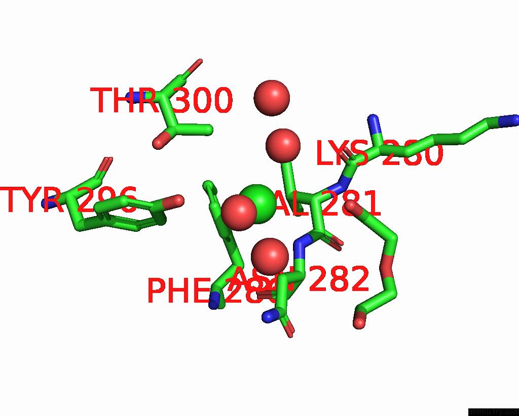
Mono view
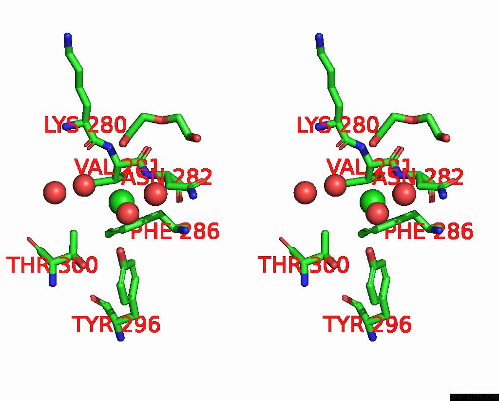
Stereo pair view
Mono view
Stereo pair view
A full contact list of Chlorine with other atoms in the Cl binding
site number 1 of High-Resolution Structure of the Siderophore Periplasmic Binding Protein Ftsb Mutant Y137A From Streptococcus Pyogenes within 5.0Å range:
|
Reference:
J.Fernandez-Perez,
J.M.M.Caaveiro,
A.Senoo,
M.Nakakido,
S.De Vega,
I.Nakagawa,
K.Tsumoto.
Conserved Binding Mechanism For Ligand Promiscuity in the Hydroxamate Siderophore Binding Protein Ftsb From Streptococcus Pyogenes Structure.
ISSN: ISSN 0969-2126
DOI: 10.1016/J.STR.2024.09.018
Page generated: Sun Jul 13 15:41:08 2025
ISSN: ISSN 0969-2126
DOI: 10.1016/J.STR.2024.09.018
Last articles
Fe in 2YXOFe in 2YRS
Fe in 2YXC
Fe in 2YNM
Fe in 2YVJ
Fe in 2YP1
Fe in 2YU2
Fe in 2YU1
Fe in 2YQB
Fe in 2YOO