Chlorine »
PDB 8ylh-8z75 »
8yu5 »
Chlorine in PDB 8yu5: The Structure of Non-Activated Thiocyanate Dehydrogenase Mutant with the H447Q Substitution From Pelomicrobium Methylotrophicum (Pmtcdh H447Q)
Protein crystallography data
The structure of The Structure of Non-Activated Thiocyanate Dehydrogenase Mutant with the H447Q Substitution From Pelomicrobium Methylotrophicum (Pmtcdh H447Q), PDB code: 8yu5
was solved by
L.A.Varfolomeeva,
N.S.Shipkov,
N.I.Dergousova,
K.M.Boyko,
T.V.Tikhonova,
V.O.Popov,
with X-Ray Crystallography technique. A brief refinement statistics is given in the table below:
| Resolution Low / High (Å) | 45.88 / 1.45 |
| Space group | P 21 21 21 |
| Cell size a, b, c (Å), α, β, γ (°) | 66.77, 96.44, 147.67, 90, 90, 90 |
| R / Rfree (%) | 15 / 17.3 |
Other elements in 8yu5:
The structure of The Structure of Non-Activated Thiocyanate Dehydrogenase Mutant with the H447Q Substitution From Pelomicrobium Methylotrophicum (Pmtcdh H447Q) also contains other interesting chemical elements:
| Copper | (Cu) | 5 atoms |
Chlorine Binding Sites:
The binding sites of Chlorine atom in the The Structure of Non-Activated Thiocyanate Dehydrogenase Mutant with the H447Q Substitution From Pelomicrobium Methylotrophicum (Pmtcdh H447Q)
(pdb code 8yu5). This binding sites where shown within
5.0 Angstroms radius around Chlorine atom.
In total only one binding site of Chlorine was determined in the The Structure of Non-Activated Thiocyanate Dehydrogenase Mutant with the H447Q Substitution From Pelomicrobium Methylotrophicum (Pmtcdh H447Q), PDB code: 8yu5:
In total only one binding site of Chlorine was determined in the The Structure of Non-Activated Thiocyanate Dehydrogenase Mutant with the H447Q Substitution From Pelomicrobium Methylotrophicum (Pmtcdh H447Q), PDB code: 8yu5:
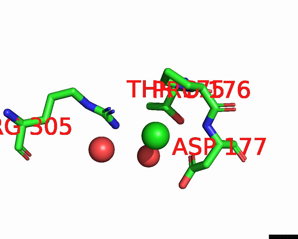
Chlorine binding site 1 out of 1 in 8yu5
Go back to
Chlorine binding site 1 out
of 1 in the The Structure of Non-Activated Thiocyanate Dehydrogenase Mutant with the H447Q Substitution From Pelomicrobium Methylotrophicum (Pmtcdh H447Q)
Mono view

Stereo pair view
Mono view
Stereo pair view
A full contact list of Chlorine with other atoms in the Cl binding
site number 1 of The Structure of Non-Activated Thiocyanate Dehydrogenase Mutant with the H447Q Substitution From Pelomicrobium Methylotrophicum (Pmtcdh H447Q) within 5.0Å range:
|
Reference:
L.A.Varfolomeeva,
N.S.Shipkov,
N.I.Dergousova,
K.M.Boyko,
T.V.Tikhonova,
V.O.Popov.
The Structure of Non-Activated Thiocyanate Dehydrogenase Mutant with the H447Q Substitution From Pelomicrobium Methylotrophicum (Pmtcdh H447Q) To Be Published.
Page generated: Sun Jul 13 15:48:02 2025
Last articles
F in 7OARF in 7OD9
F in 7OFD
F in 7O7K
F in 7OFA
F in 7OAM
F in 7O7J
F in 7O75
F in 7O73
F in 7O72