Chlorine »
PDB 9g7o-9h2d »
9gf1 »
Chlorine in PDB 9gf1: Cysteine-Less Human Galectin-3 C173S Mutant Carbohydrate Recognition Domain
Protein crystallography data
The structure of Cysteine-Less Human Galectin-3 C173S Mutant Carbohydrate Recognition Domain, PDB code: 9gf1
was solved by
J.Blaha,
Y.Dubanych,
O.Skorepa,
O.Vanek,
with X-Ray Crystallography technique. A brief refinement statistics is given in the table below:
| Resolution Low / High (Å) | 42.75 / 1.50 |
| Space group | P 21 21 21 |
| Cell size a, b, c (Å), α, β, γ (°) | 37.02, 58.047, 63.184, 90, 90, 90 |
| R / Rfree (%) | 16.1 / 18.6 |
Chlorine Binding Sites:
The binding sites of Chlorine atom in the Cysteine-Less Human Galectin-3 C173S Mutant Carbohydrate Recognition Domain
(pdb code 9gf1). This binding sites where shown within
5.0 Angstroms radius around Chlorine atom.
In total 3 binding sites of Chlorine where determined in the Cysteine-Less Human Galectin-3 C173S Mutant Carbohydrate Recognition Domain, PDB code: 9gf1:
Jump to Chlorine binding site number: 1; 2; 3;
In total 3 binding sites of Chlorine where determined in the Cysteine-Less Human Galectin-3 C173S Mutant Carbohydrate Recognition Domain, PDB code: 9gf1:
Jump to Chlorine binding site number: 1; 2; 3;
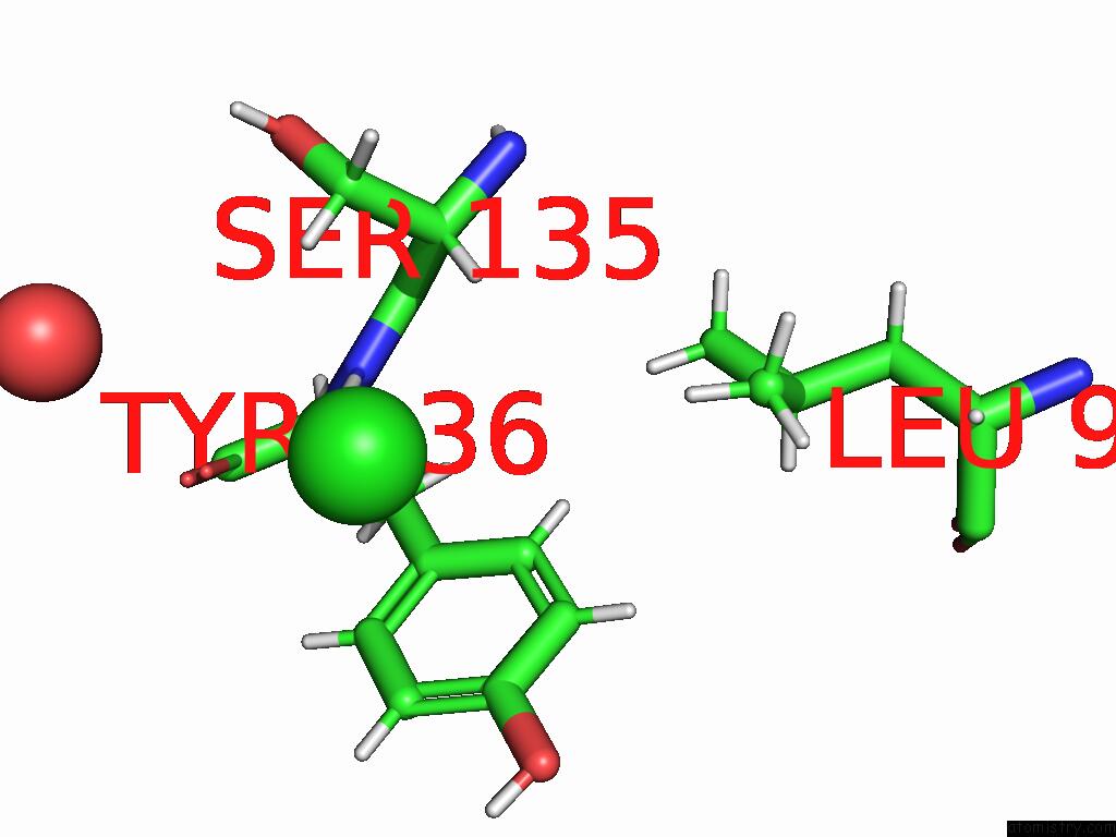
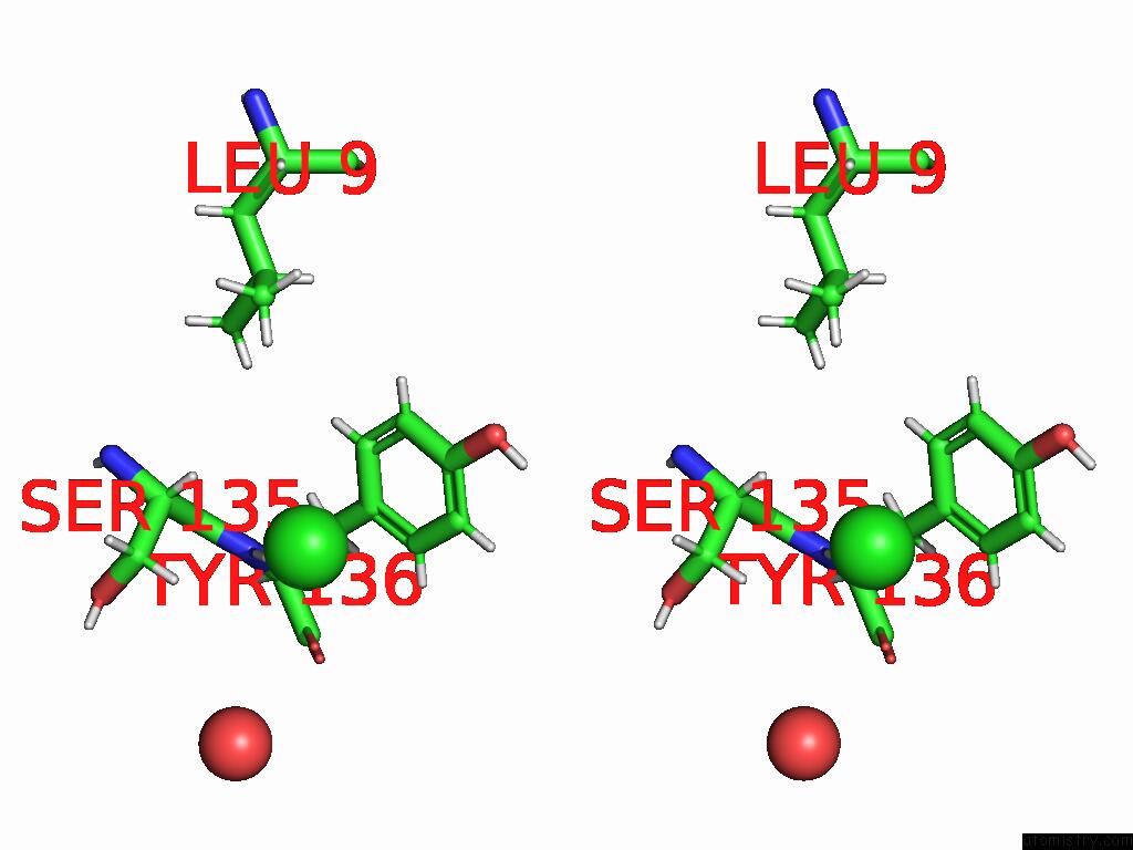
Chlorine binding site 1 out of 3 in 9gf1
Go back to
Chlorine binding site 1 out
of 3 in the Cysteine-Less Human Galectin-3 C173S Mutant Carbohydrate Recognition Domain
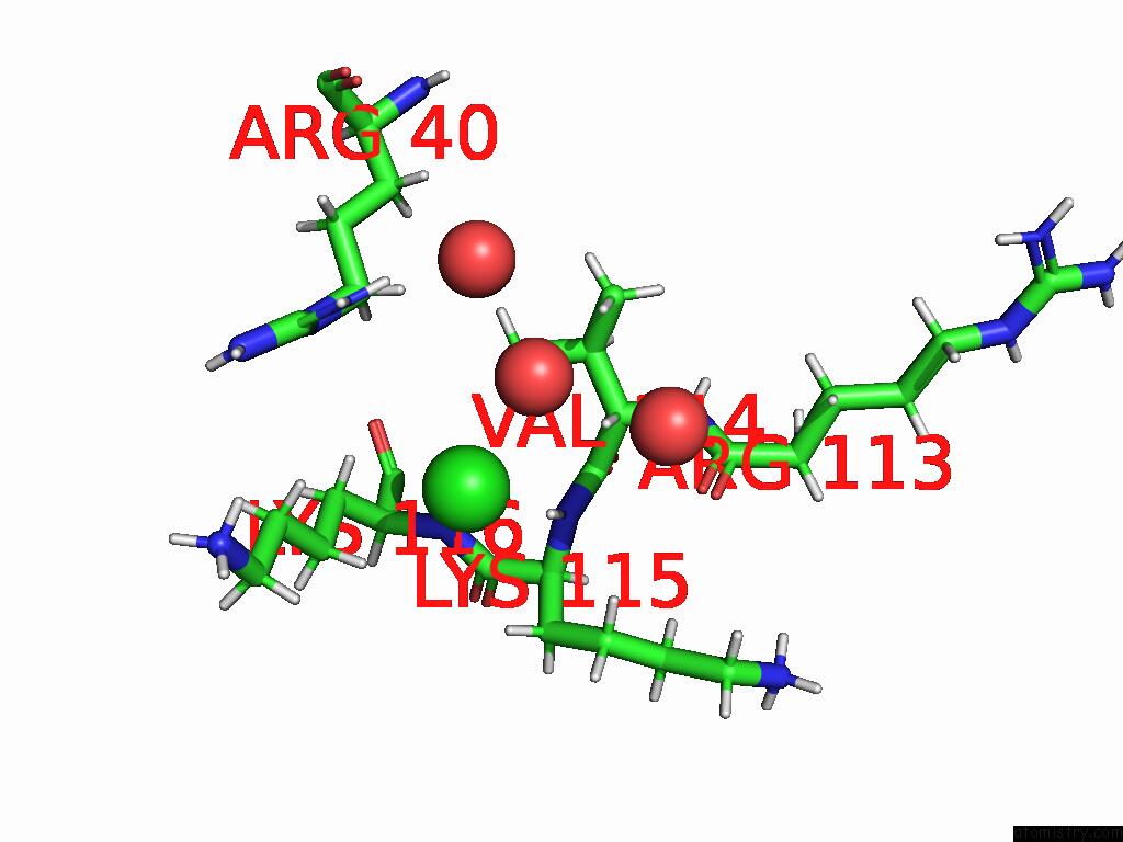
Mono view
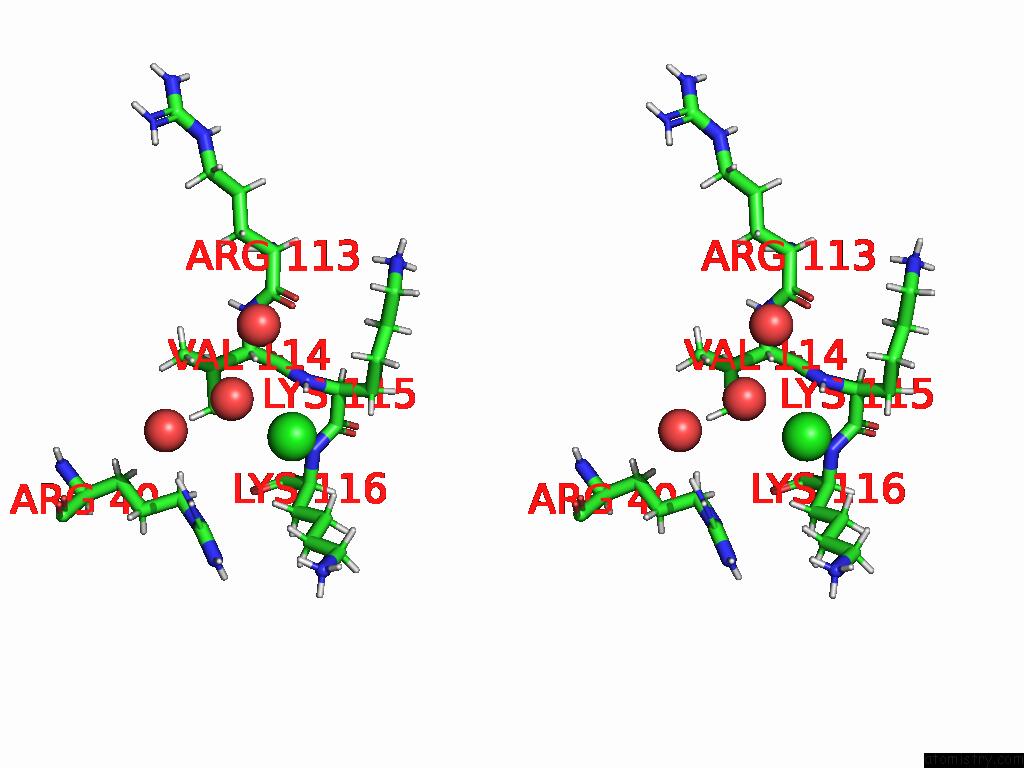
Stereo pair view
Mono view
Stereo pair view
A full contact list of Chlorine with other atoms in the Cl binding
site number 1 of Cysteine-Less Human Galectin-3 C173S Mutant Carbohydrate Recognition Domain within 5.0Å range:
|
Chlorine binding site 2 out of 3 in 9gf1
Go back to
Chlorine binding site 2 out
of 3 in the Cysteine-Less Human Galectin-3 C173S Mutant Carbohydrate Recognition Domain
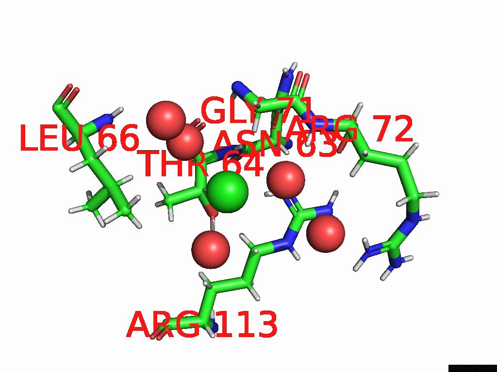
Mono view
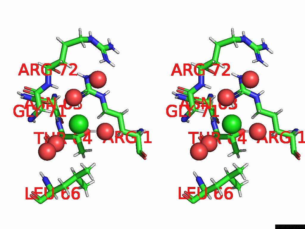
Stereo pair view
Mono view
Stereo pair view
A full contact list of Chlorine with other atoms in the Cl binding
site number 2 of Cysteine-Less Human Galectin-3 C173S Mutant Carbohydrate Recognition Domain within 5.0Å range:
|
Chlorine binding site 3 out of 3 in 9gf1
Go back to
Chlorine binding site 3 out
of 3 in the Cysteine-Less Human Galectin-3 C173S Mutant Carbohydrate Recognition Domain
Mono view
Stereo pair view
Mono view
Stereo pair view
A full contact list of Chlorine with other atoms in the Cl binding
site number 3 of Cysteine-Less Human Galectin-3 C173S Mutant Carbohydrate Recognition Domain within 5.0Å range:
|
Reference:
J.Blaha,
Y.Dubanych,
O.Skorepa,
O.Vanek.
Galectin-3 Carbohydrate Recognition Is Solely Responsible For Its Binding to the Nk Cell Activation Receptor NKP30. To Be Published.
Page generated: Tue Aug 26 20:07:19 2025
Last articles
Zn in 9QM9Zn in 9S44
Zn in 9OFE
Zn in 9OFC
Zn in 9OFD
Zn in 9OF1
Zn in 9OFB
Zn in 9N0J
Zn in 9M5X
Zn in 9LGI