Chlorine »
PDB 9lvx-9oa8 »
9mh2 »
Chlorine in PDB 9mh2: Crystal Structure of Purine Nucleoside Phosphorylase From Trichomonas Vaginalis (Adenosine and Glycine Complex)
Protein crystallography data
The structure of Crystal Structure of Purine Nucleoside Phosphorylase From Trichomonas Vaginalis (Adenosine and Glycine Complex), PDB code: 9mh2
was solved by
Seattle Structural Genomics Center For Infectious Disease,
Seattlestructural Genomics Center For Infectious Disease (Ssgcid),
with X-Ray Crystallography technique. A brief refinement statistics is given in the table below:
| Resolution Low / High (Å) | 46.52 / 1.35 |
| Space group | H 3 2 |
| Cell size a, b, c (Å), α, β, γ (°) | 93.033, 93.033, 130.449, 90, 90, 120 |
| R / Rfree (%) | 14.2 / 16.8 |
Chlorine Binding Sites:
The binding sites of Chlorine atom in the Crystal Structure of Purine Nucleoside Phosphorylase From Trichomonas Vaginalis (Adenosine and Glycine Complex)
(pdb code 9mh2). This binding sites where shown within
5.0 Angstroms radius around Chlorine atom.
In total only one binding site of Chlorine was determined in the Crystal Structure of Purine Nucleoside Phosphorylase From Trichomonas Vaginalis (Adenosine and Glycine Complex), PDB code: 9mh2:
In total only one binding site of Chlorine was determined in the Crystal Structure of Purine Nucleoside Phosphorylase From Trichomonas Vaginalis (Adenosine and Glycine Complex), PDB code: 9mh2:
Chlorine binding site 1 out of 1 in 9mh2
Go back to
Chlorine binding site 1 out
of 1 in the Crystal Structure of Purine Nucleoside Phosphorylase From Trichomonas Vaginalis (Adenosine and Glycine Complex)
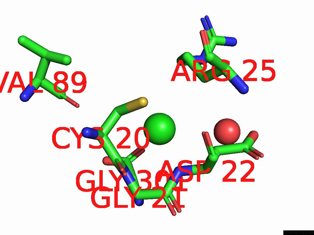
Mono view
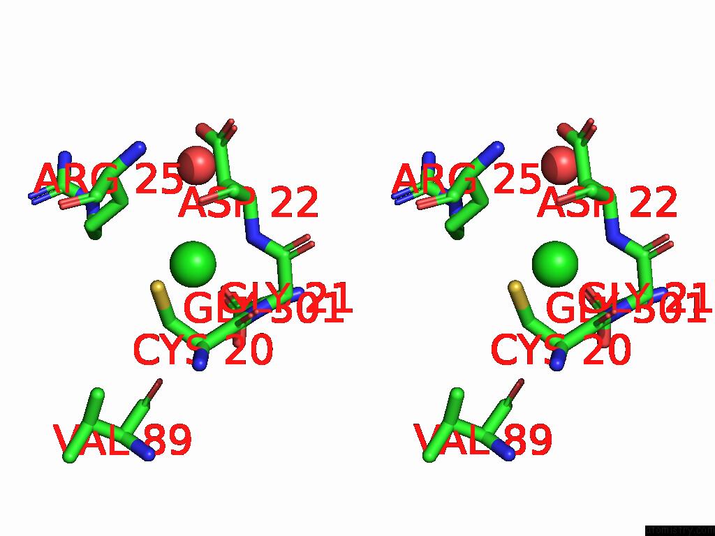
Stereo pair view
Mono view
Stereo pair view
A full contact list of Chlorine with other atoms in the Cl binding
site number 1 of Crystal Structure of Purine Nucleoside Phosphorylase From Trichomonas Vaginalis (Adenosine and Glycine Complex) within 5.0Å range:
|
Reference:
S.Seibold,
L.Liu,
S.Lovell,
K.P.Battaile.
Crystal Structure of Purine Nucleoside Phosphorylase From Trichomonas Vaginalis (Adenosine and Glycine Complex) To Be Published.
Page generated: Sun Jul 13 17:14:04 2025
Last articles
F in 7N4QF in 7N4N
F in 7N2A
F in 7MXY
F in 7MYY
F in 7N13
F in 7MYU
F in 7MYR
F in 7MYO
F in 7MXN