Chlorine »
PDB 9orv-9v6z »
9qcg »
Chlorine in PDB 9qcg: Crystal Structure of Methanopyrus Kandleri Malate Dehydrogenase Mutant 4 at Room Temperature
Enzymatic activity of Crystal Structure of Methanopyrus Kandleri Malate Dehydrogenase Mutant 4 at Room Temperature
All present enzymatic activity of Crystal Structure of Methanopyrus Kandleri Malate Dehydrogenase Mutant 4 at Room Temperature:
1.1.1.299;
1.1.1.299;
Protein crystallography data
The structure of Crystal Structure of Methanopyrus Kandleri Malate Dehydrogenase Mutant 4 at Room Temperature, PDB code: 9qcg
was solved by
S.Coquille,
D.Madern,
with X-Ray Crystallography technique. A brief refinement statistics is given in the table below:
| Resolution Low / High (Å) | 17.90 / 2.39 |
| Space group | P 41 21 2 |
| Cell size a, b, c (Å), α, β, γ (°) | 75.305, 75.305, 270.065, 90, 90, 90 |
| R / Rfree (%) | 26.3 / 30.9 |
Other elements in 9qcg:
The structure of Crystal Structure of Methanopyrus Kandleri Malate Dehydrogenase Mutant 4 at Room Temperature also contains other interesting chemical elements:
| Potassium | (K) | 2 atoms |
Chlorine Binding Sites:
The binding sites of Chlorine atom in the Crystal Structure of Methanopyrus Kandleri Malate Dehydrogenase Mutant 4 at Room Temperature
(pdb code 9qcg). This binding sites where shown within
5.0 Angstroms radius around Chlorine atom.
In total 3 binding sites of Chlorine where determined in the Crystal Structure of Methanopyrus Kandleri Malate Dehydrogenase Mutant 4 at Room Temperature, PDB code: 9qcg:
Jump to Chlorine binding site number: 1; 2; 3;
In total 3 binding sites of Chlorine where determined in the Crystal Structure of Methanopyrus Kandleri Malate Dehydrogenase Mutant 4 at Room Temperature, PDB code: 9qcg:
Jump to Chlorine binding site number: 1; 2; 3;
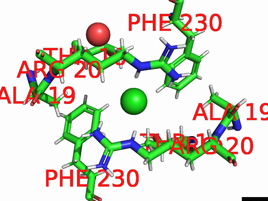
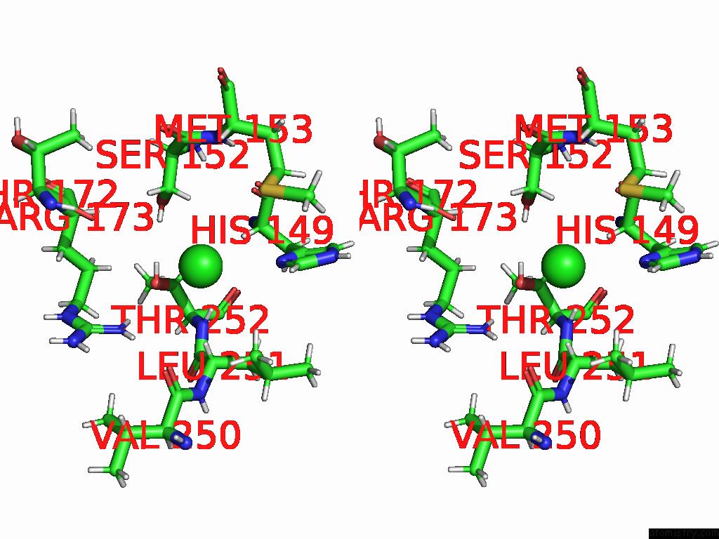
Chlorine binding site 1 out of 3 in 9qcg
Go back to
Chlorine binding site 1 out
of 3 in the Crystal Structure of Methanopyrus Kandleri Malate Dehydrogenase Mutant 4 at Room Temperature
Mono view
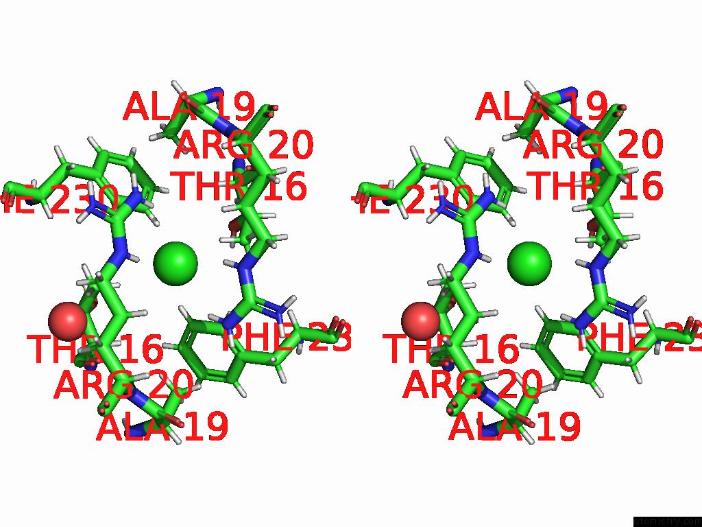
Stereo pair view
Mono view
Stereo pair view
A full contact list of Chlorine with other atoms in the Cl binding
site number 1 of Crystal Structure of Methanopyrus Kandleri Malate Dehydrogenase Mutant 4 at Room Temperature within 5.0Å range:
|
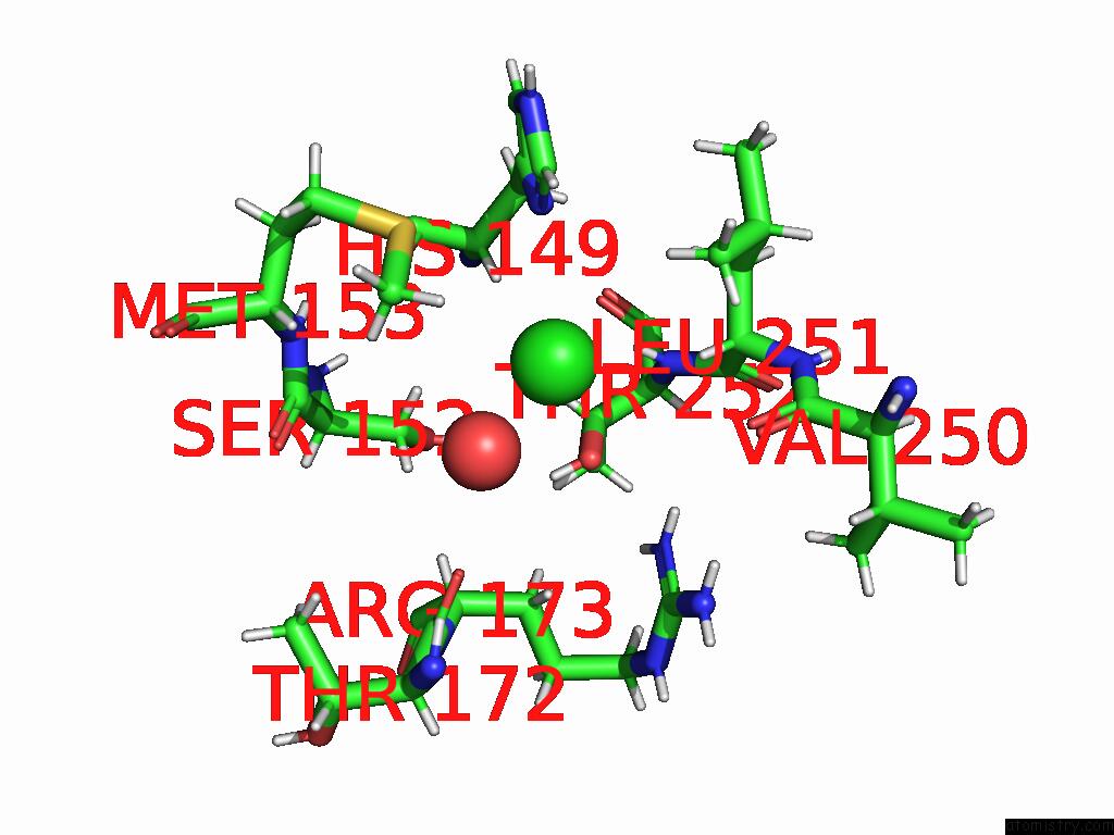
Chlorine binding site 2 out of 3 in 9qcg
Go back to
Chlorine binding site 2 out
of 3 in the Crystal Structure of Methanopyrus Kandleri Malate Dehydrogenase Mutant 4 at Room Temperature
Mono view

Stereo pair view
Mono view
Stereo pair view
A full contact list of Chlorine with other atoms in the Cl binding
site number 2 of Crystal Structure of Methanopyrus Kandleri Malate Dehydrogenase Mutant 4 at Room Temperature within 5.0Å range:
|
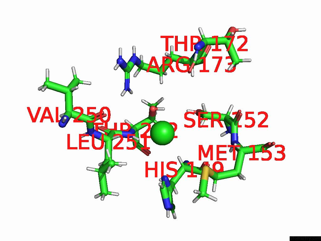
Chlorine binding site 3 out of 3 in 9qcg
Go back to
Chlorine binding site 3 out
of 3 in the Crystal Structure of Methanopyrus Kandleri Malate Dehydrogenase Mutant 4 at Room Temperature
Mono view
Stereo pair view
Mono view
Stereo pair view
A full contact list of Chlorine with other atoms in the Cl binding
site number 3 of Crystal Structure of Methanopyrus Kandleri Malate Dehydrogenase Mutant 4 at Room Temperature within 5.0Å range:
|
Reference:
S.Coquille,
C.S.Pereira,
J.Roche,
G.Santoni,
S.Engilberge,
C.Brochier-Armanet,
E.Girard,
F.Sterpone,
D.Madern.
Allostery and Evolution: A Molecular Journey Through the Structural and Dynamical Landscape of An Enzyme Super Family. Mol.Biol.Evol. V. 42 2025.
ISSN: ESSN 1537-1719
PubMed: 39834309
DOI: 10.1093/MOLBEV/MSAE265
Page generated: Sun Jul 13 17:20:05 2025
ISSN: ESSN 1537-1719
PubMed: 39834309
DOI: 10.1093/MOLBEV/MSAE265
Last articles
Cu in 1YAICu in 1YAZ
Cu in 1XTL
Cu in 1Y3J
Cu in 1XME
Cu in 1XTM
Cu in 1XSO
Cu in 1XB3
Cu in 1XB6
Cu in 1X9R