Chlorine »
PDB 162l-1ag9 »
1a2w »
Chlorine in PDB 1a2w: Crystal Structure of A 3D Domain-Swapped Dimer of Bovine Pancreatic Ribonuclease A
Enzymatic activity of Crystal Structure of A 3D Domain-Swapped Dimer of Bovine Pancreatic Ribonuclease A
All present enzymatic activity of Crystal Structure of A 3D Domain-Swapped Dimer of Bovine Pancreatic Ribonuclease A:
3.1.27.5;
3.1.27.5;
Protein crystallography data
The structure of Crystal Structure of A 3D Domain-Swapped Dimer of Bovine Pancreatic Ribonuclease A, PDB code: 1a2w
was solved by
Y.Liu,
P.J.Hart,
M.P.Schlunegger,
D.S.Eisenberg,
with X-Ray Crystallography technique. A brief refinement statistics is given in the table below:
| Resolution Low / High (Å) | 10.00 / 2.10 |
| Space group | P 32 |
| Cell size a, b, c (Å), α, β, γ (°) | 57.000, 57.000, 81.410, 90.00, 90.00, 120.00 |
| R / Rfree (%) | 19.2 / 25.6 |
Chlorine Binding Sites:
The binding sites of Chlorine atom in the Crystal Structure of A 3D Domain-Swapped Dimer of Bovine Pancreatic Ribonuclease A
(pdb code 1a2w). This binding sites where shown within
5.0 Angstroms radius around Chlorine atom.
In total 2 binding sites of Chlorine where determined in the Crystal Structure of A 3D Domain-Swapped Dimer of Bovine Pancreatic Ribonuclease A, PDB code: 1a2w:
Jump to Chlorine binding site number: 1; 2;
In total 2 binding sites of Chlorine where determined in the Crystal Structure of A 3D Domain-Swapped Dimer of Bovine Pancreatic Ribonuclease A, PDB code: 1a2w:
Jump to Chlorine binding site number: 1; 2;
Chlorine binding site 1 out of 2 in 1a2w
Go back to
Chlorine binding site 1 out
of 2 in the Crystal Structure of A 3D Domain-Swapped Dimer of Bovine Pancreatic Ribonuclease A
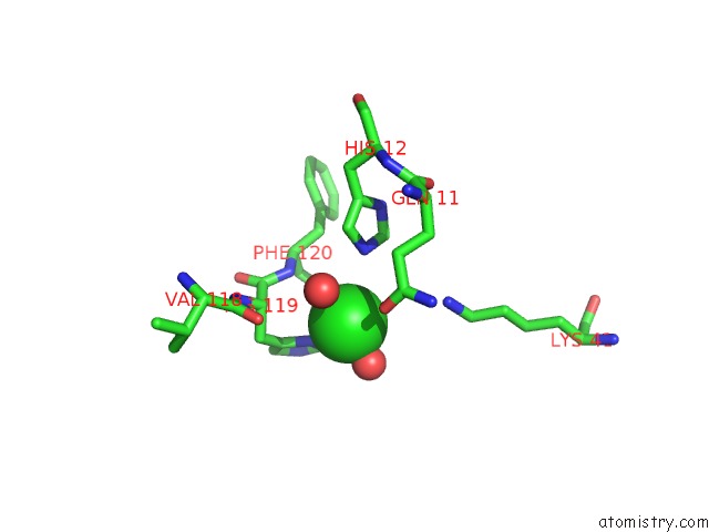
Mono view
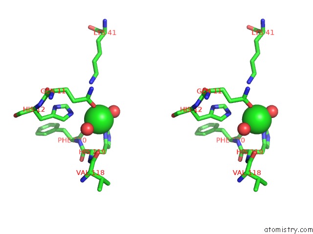
Stereo pair view
Mono view
Stereo pair view
A full contact list of Chlorine with other atoms in the Cl binding
site number 1 of Crystal Structure of A 3D Domain-Swapped Dimer of Bovine Pancreatic Ribonuclease A within 5.0Å range:
|
Chlorine binding site 2 out of 2 in 1a2w
Go back to
Chlorine binding site 2 out
of 2 in the Crystal Structure of A 3D Domain-Swapped Dimer of Bovine Pancreatic Ribonuclease A
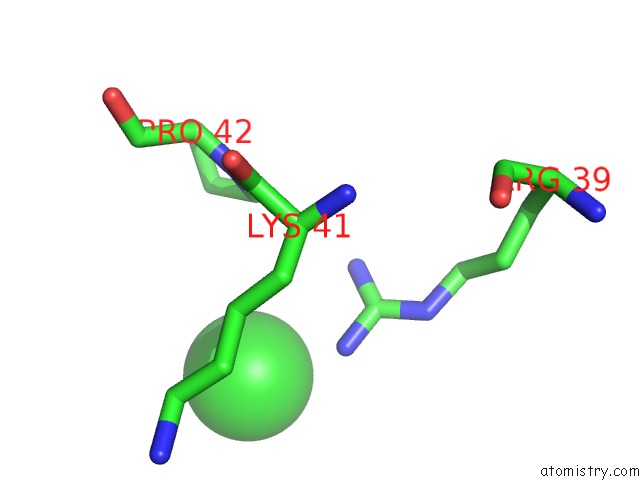
Mono view
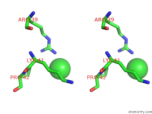
Stereo pair view
Mono view
Stereo pair view
A full contact list of Chlorine with other atoms in the Cl binding
site number 2 of Crystal Structure of A 3D Domain-Swapped Dimer of Bovine Pancreatic Ribonuclease A within 5.0Å range:
|
Reference:
Y.Liu,
P.J.Hart,
M.P.Schlunegger,
D.Eisenberg.
The Crystal Structure of A 3D Domain-Swapped Dimer of Rnase A at A 2.1-A Resolution. Proc.Natl.Acad.Sci.Usa V. 95 3437 1998.
ISSN: ISSN 0027-8424
PubMed: 9520384
DOI: 10.1073/PNAS.95.7.3437
Page generated: Thu Jul 10 16:14:41 2025
ISSN: ISSN 0027-8424
PubMed: 9520384
DOI: 10.1073/PNAS.95.7.3437
Last articles
Mg in 4NJIMg in 4NIA
Mg in 4NH1
Mg in 4NH0
Mg in 4NI5
Mg in 4NG6
Mg in 4NG4
Mg in 4NFJ
Mg in 4NFI
Mg in 4NFL