Chlorine »
PDB 1cu6-1dhi »
1cx7 »
Chlorine in PDB 1cx7: T4 Lysozyme Methionine Core Mutant
Enzymatic activity of T4 Lysozyme Methionine Core Mutant
All present enzymatic activity of T4 Lysozyme Methionine Core Mutant:
3.2.1.17;
3.2.1.17;
Protein crystallography data
The structure of T4 Lysozyme Methionine Core Mutant, PDB code: 1cx7
was solved by
N.C.Gassner,
W.A.Baase,
J.Lindstrom,
J.Lu,
B.W.Matthews,
with X-Ray Crystallography technique. A brief refinement statistics is given in the table below:
| Resolution Low / High (Å) | 30.00 / 1.94 |
| Space group | P 32 2 1 |
| Cell size a, b, c (Å), α, β, γ (°) | 61.310, 61.310, 96.490, 90.00, 90.00, 120.00 |
| R / Rfree (%) | n/a / n/a |
Chlorine Binding Sites:
The binding sites of Chlorine atom in the T4 Lysozyme Methionine Core Mutant
(pdb code 1cx7). This binding sites where shown within
5.0 Angstroms radius around Chlorine atom.
In total 2 binding sites of Chlorine where determined in the T4 Lysozyme Methionine Core Mutant, PDB code: 1cx7:
Jump to Chlorine binding site number: 1; 2;
In total 2 binding sites of Chlorine where determined in the T4 Lysozyme Methionine Core Mutant, PDB code: 1cx7:
Jump to Chlorine binding site number: 1; 2;
Chlorine binding site 1 out of 2 in 1cx7
Go back to
Chlorine binding site 1 out
of 2 in the T4 Lysozyme Methionine Core Mutant
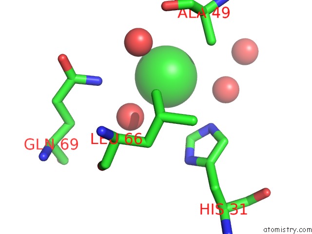
Mono view
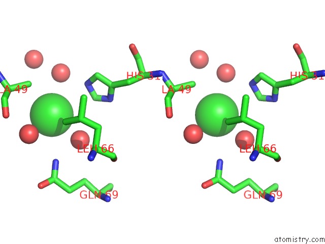
Stereo pair view
Mono view
Stereo pair view
A full contact list of Chlorine with other atoms in the Cl binding
site number 1 of T4 Lysozyme Methionine Core Mutant within 5.0Å range:
|
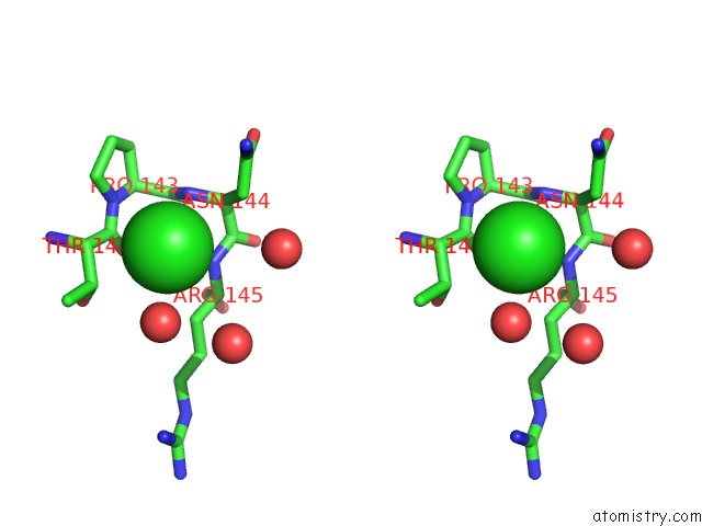
Chlorine binding site 2 out of 2 in 1cx7
Go back to
Chlorine binding site 2 out
of 2 in the T4 Lysozyme Methionine Core Mutant
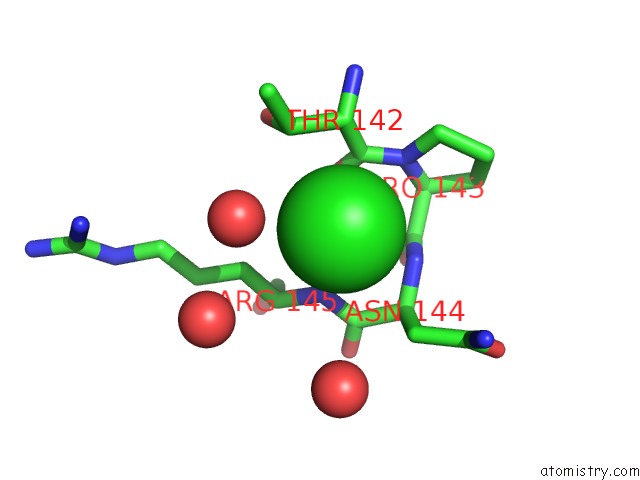
Mono view
Stereo pair view
Mono view
Stereo pair view
A full contact list of Chlorine with other atoms in the Cl binding
site number 2 of T4 Lysozyme Methionine Core Mutant within 5.0Å range:
|
Reference:
N.C.Gassner,
B.W.Matthews.
Use of Differentially Substituted Selenomethionine Proteins in X-Ray Structure Determination. Acta Crystallogr.,Sect.D V. 55 1967 1999.
ISSN: ISSN 0907-4449
PubMed: 10666571
DOI: 10.1021/BI9915519
Page generated: Thu Jul 10 16:36:39 2025
ISSN: ISSN 0907-4449
PubMed: 10666571
DOI: 10.1021/BI9915519
Last articles
I in 5W4PI in 5WKG
I in 5WL1
I in 5W4O
I in 5W4Q
I in 5W4I
I in 5U84
I in 5W1H
I in 5W1I
I in 5W0M