Chlorine »
PDB 1cu6-1dhi »
1dgm »
Chlorine in PDB 1dgm: Crystal Structure of Adenosine Kinase From Toxoplasma Gondii
Enzymatic activity of Crystal Structure of Adenosine Kinase From Toxoplasma Gondii
All present enzymatic activity of Crystal Structure of Adenosine Kinase From Toxoplasma Gondii:
2.7.1.20;
2.7.1.20;
Protein crystallography data
The structure of Crystal Structure of Adenosine Kinase From Toxoplasma Gondii, PDB code: 1dgm
was solved by
W.J.Cook,
L.J.Delucas,
D.Chattopadhyay,
with X-Ray Crystallography technique. A brief refinement statistics is given in the table below:
| Resolution Low / High (Å) | 46.78 / 1.80 |
| Space group | P 1 21 1 |
| Cell size a, b, c (Å), α, β, γ (°) | 47.550, 68.750, 57.290, 90.00, 100.30, 90.00 |
| R / Rfree (%) | 21.4 / 23.3 |
Other elements in 1dgm:
The structure of Crystal Structure of Adenosine Kinase From Toxoplasma Gondii also contains other interesting chemical elements:
| Magnesium | (Mg) | 1 atom |
Chlorine Binding Sites:
The binding sites of Chlorine atom in the Crystal Structure of Adenosine Kinase From Toxoplasma Gondii
(pdb code 1dgm). This binding sites where shown within
5.0 Angstroms radius around Chlorine atom.
In total only one binding site of Chlorine was determined in the Crystal Structure of Adenosine Kinase From Toxoplasma Gondii, PDB code: 1dgm:
In total only one binding site of Chlorine was determined in the Crystal Structure of Adenosine Kinase From Toxoplasma Gondii, PDB code: 1dgm:
Chlorine binding site 1 out of 1 in 1dgm
Go back to
Chlorine binding site 1 out
of 1 in the Crystal Structure of Adenosine Kinase From Toxoplasma Gondii
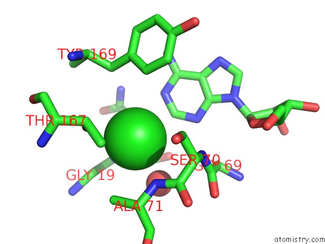
Mono view
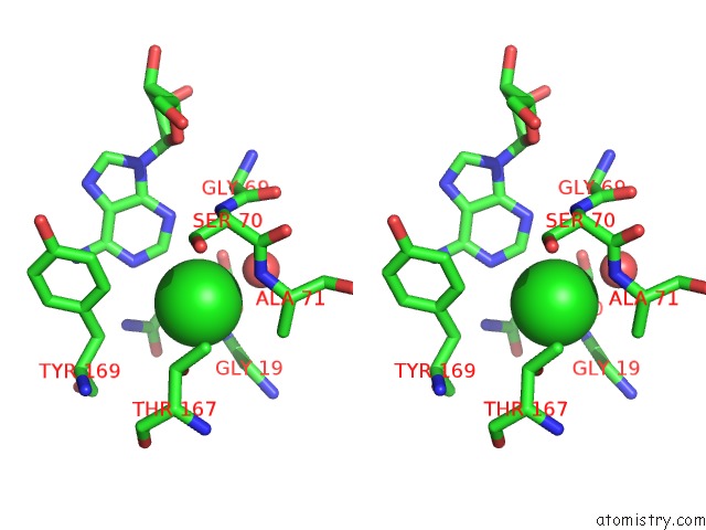
Stereo pair view
Mono view
Stereo pair view
A full contact list of Chlorine with other atoms in the Cl binding
site number 1 of Crystal Structure of Adenosine Kinase From Toxoplasma Gondii within 5.0Å range:
|
Reference:
W.J.Cook,
L.J.Delucas,
D.Chattopadhyay.
Crystal Structure of Adenosine Kinase From Toxoplasma Gondii at 1.8 A Resolution. Protein Sci. V. 9 704 2000.
ISSN: ISSN 0961-8368
PubMed: 10794412
Page generated: Thu Jul 10 16:38:54 2025
ISSN: ISSN 0961-8368
PubMed: 10794412
Last articles
La in 5YTQLa in 6IP9
La in 1DJG
La in 6DAM
La in 5KKB
La in 2OQR
La in 2RPV
La in 5KIJ
La in 2K0J
La in 2I18