Chlorine »
PDB 1dhj-1e2y »
1diq »
Chlorine in PDB 1diq: Crystal Structure of P-Cresol Methylhydroxylase with Substrate Bound
Enzymatic activity of Crystal Structure of P-Cresol Methylhydroxylase with Substrate Bound
All present enzymatic activity of Crystal Structure of P-Cresol Methylhydroxylase with Substrate Bound:
1.17.99.1;
1.17.99.1;
Protein crystallography data
The structure of Crystal Structure of P-Cresol Methylhydroxylase with Substrate Bound, PDB code: 1diq
was solved by
L.M.Cunane,
Z.W.Chen,
N.Shamala,
F.S.Mathews,
C.S.Cronin,
W.S.Mcintire,
with X-Ray Crystallography technique. A brief refinement statistics is given in the table below:
| Resolution Low / High (Å) | 25.00 / 2.75 |
| Space group | P 21 21 21 |
| Cell size a, b, c (Å), α, β, γ (°) | 139.100, 130.500, 74.400, 90.00, 90.00, 90.00 |
| R / Rfree (%) | 13.3 / 18.3 |
Other elements in 1diq:
The structure of Crystal Structure of P-Cresol Methylhydroxylase with Substrate Bound also contains other interesting chemical elements:
| Iron | (Fe) | 2 atoms |
Chlorine Binding Sites:
The binding sites of Chlorine atom in the Crystal Structure of P-Cresol Methylhydroxylase with Substrate Bound
(pdb code 1diq). This binding sites where shown within
5.0 Angstroms radius around Chlorine atom.
In total 2 binding sites of Chlorine where determined in the Crystal Structure of P-Cresol Methylhydroxylase with Substrate Bound, PDB code: 1diq:
Jump to Chlorine binding site number: 1; 2;
In total 2 binding sites of Chlorine where determined in the Crystal Structure of P-Cresol Methylhydroxylase with Substrate Bound, PDB code: 1diq:
Jump to Chlorine binding site number: 1; 2;
Chlorine binding site 1 out of 2 in 1diq
Go back to
Chlorine binding site 1 out
of 2 in the Crystal Structure of P-Cresol Methylhydroxylase with Substrate Bound
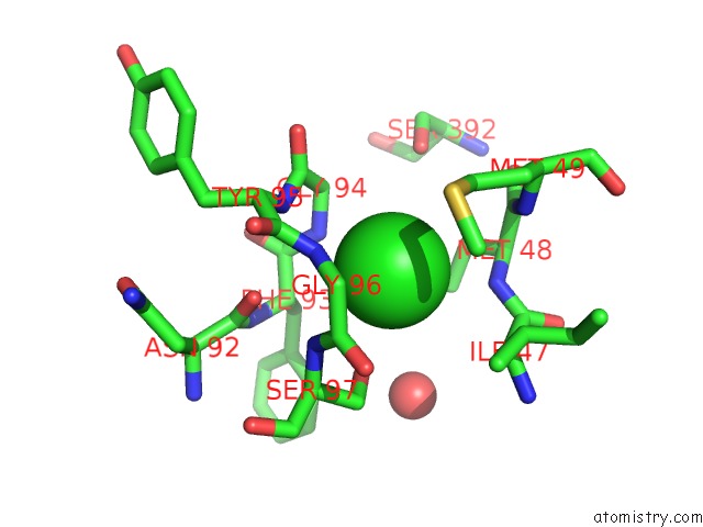
Mono view
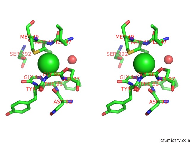
Stereo pair view
Mono view
Stereo pair view
A full contact list of Chlorine with other atoms in the Cl binding
site number 1 of Crystal Structure of P-Cresol Methylhydroxylase with Substrate Bound within 5.0Å range:
|
Chlorine binding site 2 out of 2 in 1diq
Go back to
Chlorine binding site 2 out
of 2 in the Crystal Structure of P-Cresol Methylhydroxylase with Substrate Bound
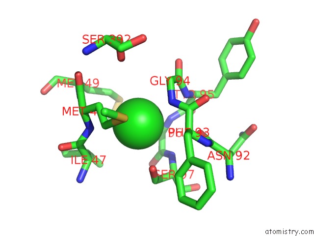
Mono view
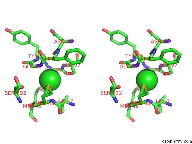
Stereo pair view
Mono view
Stereo pair view
A full contact list of Chlorine with other atoms in the Cl binding
site number 2 of Crystal Structure of P-Cresol Methylhydroxylase with Substrate Bound within 5.0Å range:
|
Reference:
L.M.Cunane,
Z.W.Chen,
N.Shamala,
F.S.Mathews,
C.N.Cronin,
W.S.Mcintire.
Structures of the Flavocytochrome P-Cresol Methylhydroxylase and Its Enzyme-Substrate Complex: Gated Substrate Entry and Proton Relays Support the Proposed Catalytic Mechanism. J.Mol.Biol. V. 295 357 2000.
ISSN: ISSN 0022-2836
PubMed: 10623531
DOI: 10.1006/JMBI.1999.3290
Page generated: Thu Jul 10 16:39:13 2025
ISSN: ISSN 0022-2836
PubMed: 10623531
DOI: 10.1006/JMBI.1999.3290
Last articles
K in 8CSRK in 8CGD
K in 8CSQ
K in 8CSP
K in 8CRD
K in 8CLM
K in 8CJ7
K in 8CGU
K in 8CGR
K in 8CGJ