Chlorine »
PDB 1e3a-1exv »
1e66 »
Chlorine in PDB 1e66: Structure of Acetylcholinesterase Complexed with (-)-Huprine X at 2.1A Resolution
Enzymatic activity of Structure of Acetylcholinesterase Complexed with (-)-Huprine X at 2.1A Resolution
All present enzymatic activity of Structure of Acetylcholinesterase Complexed with (-)-Huprine X at 2.1A Resolution:
3.1.1.7;
3.1.1.7;
Protein crystallography data
The structure of Structure of Acetylcholinesterase Complexed with (-)-Huprine X at 2.1A Resolution, PDB code: 1e66
was solved by
H.Dvir,
M.Harel,
I.Silman,
J.L.Sussman,
with X-Ray Crystallography technique. A brief refinement statistics is given in the table below:
| Resolution Low / High (Å) | 29.42 / 2.10 |
| Space group | P 31 2 1 |
| Cell size a, b, c (Å), α, β, γ (°) | 112.335, 112.335, 138.165, 90.00, 90.00, 120.00 |
| R / Rfree (%) | 17.7 / 20.5 |
Chlorine Binding Sites:
The binding sites of Chlorine atom in the Structure of Acetylcholinesterase Complexed with (-)-Huprine X at 2.1A Resolution
(pdb code 1e66). This binding sites where shown within
5.0 Angstroms radius around Chlorine atom.
In total only one binding site of Chlorine was determined in the Structure of Acetylcholinesterase Complexed with (-)-Huprine X at 2.1A Resolution, PDB code: 1e66:
In total only one binding site of Chlorine was determined in the Structure of Acetylcholinesterase Complexed with (-)-Huprine X at 2.1A Resolution, PDB code: 1e66:
Chlorine binding site 1 out of 1 in 1e66
Go back to
Chlorine binding site 1 out
of 1 in the Structure of Acetylcholinesterase Complexed with (-)-Huprine X at 2.1A Resolution
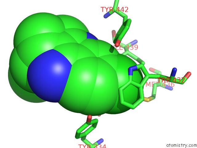
Mono view
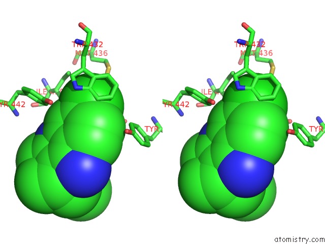
Stereo pair view
Mono view
Stereo pair view
A full contact list of Chlorine with other atoms in the Cl binding
site number 1 of Structure of Acetylcholinesterase Complexed with (-)-Huprine X at 2.1A Resolution within 5.0Å range:
|
Reference:
H.Dvir,
D.M.Wong,
M.Harel,
X.Barril,
M.Orozco,
F.J.Luque,
D.Munoz-Torrero,
P.Camps,
T.L.Rosenberry,
I.Silman,
J.L.Sussman.
3D Structure of Torpedo Californica Acetylcholinesterase Complexed with Huprine X at 2. 1 A Resolution: Kinetic and Molecular Dynamic Correlates. Biochemistry V. 41 2970 2002.
ISSN: ISSN 0006-2960
PubMed: 11863435
DOI: 10.1021/BI011652I
Page generated: Thu Jul 10 16:45:04 2025
ISSN: ISSN 0006-2960
PubMed: 11863435
DOI: 10.1021/BI011652I
Last articles
K in 4RBQK in 4R8I
K in 4R4V
K in 4R7J
K in 4R1L
K in 4R47
K in 4R45
K in 4R33
K in 4R2C
K in 4QXG