Chlorine »
PDB 1e3a-1exv »
1evr »
Chlorine in PDB 1evr: The Structure of the Resorcinol/Insulin R6 Hexamer
Protein crystallography data
The structure of The Structure of the Resorcinol/Insulin R6 Hexamer, PDB code: 1evr
was solved by
G.D.Smith,
E.Ciszak,
L.A.Magrum,
W.A.Pangborn,
R.H.Blessing,
with X-Ray Crystallography technique. A brief refinement statistics is given in the table below:
| Resolution Low / High (Å) | 44.75 / 1.90 |
| Space group | P 1 21 1 |
| Cell size a, b, c (Å), α, β, γ (°) | 61.350, 61.926, 47.805, 90.00, 110.61, 90.00 |
| R / Rfree (%) | 17.9 / 21.8 |
Other elements in 1evr:
The structure of The Structure of the Resorcinol/Insulin R6 Hexamer also contains other interesting chemical elements:
| Zinc | (Zn) | 2 atoms |
| Sodium | (Na) | 1 atom |
Chlorine Binding Sites:
The binding sites of Chlorine atom in the The Structure of the Resorcinol/Insulin R6 Hexamer
(pdb code 1evr). This binding sites where shown within
5.0 Angstroms radius around Chlorine atom.
In total 2 binding sites of Chlorine where determined in the The Structure of the Resorcinol/Insulin R6 Hexamer, PDB code: 1evr:
Jump to Chlorine binding site number: 1; 2;
In total 2 binding sites of Chlorine where determined in the The Structure of the Resorcinol/Insulin R6 Hexamer, PDB code: 1evr:
Jump to Chlorine binding site number: 1; 2;
Chlorine binding site 1 out of 2 in 1evr
Go back to
Chlorine binding site 1 out
of 2 in the The Structure of the Resorcinol/Insulin R6 Hexamer
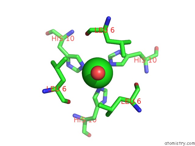
Mono view
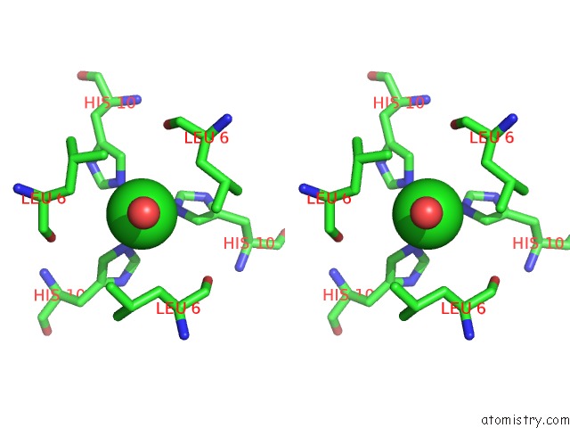
Stereo pair view
Mono view
Stereo pair view
A full contact list of Chlorine with other atoms in the Cl binding
site number 1 of The Structure of the Resorcinol/Insulin R6 Hexamer within 5.0Å range:
|
Chlorine binding site 2 out of 2 in 1evr
Go back to
Chlorine binding site 2 out
of 2 in the The Structure of the Resorcinol/Insulin R6 Hexamer
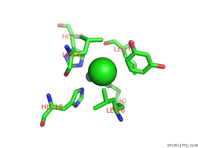
Mono view
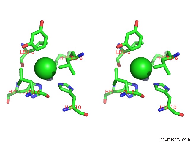
Stereo pair view
Mono view
Stereo pair view
A full contact list of Chlorine with other atoms in the Cl binding
site number 2 of The Structure of the Resorcinol/Insulin R6 Hexamer within 5.0Å range:
|
Reference:
G.D.Smith,
E.Ciszak,
L.A.Magrum,
W.A.Pangborn,
R.H.Blessing.
R6 Hexameric Insulin Complexed with M-Cresol or Resorcinol. Acta Crystallogr.,Sect.D V. 56 1541 2000.
ISSN: ISSN 0907-4449
PubMed: 11092919
DOI: 10.1107/S0907444900012749
Page generated: Thu Jul 10 16:49:21 2025
ISSN: ISSN 0907-4449
PubMed: 11092919
DOI: 10.1107/S0907444900012749
Last articles
K in 7E20K in 7DVQ
K in 7D5E
K in 7DMR
K in 7DIF
K in 7COW
K in 7D5D
K in 7D99
K in 7D52
K in 7D23