Chlorine »
PDB 1go6-1hav »
1h0r »
Chlorine in PDB 1h0r: Type II Dehydroquinase From Mycobacterium Tuberculosis Complexed with 2,3-Anhydro-Quinic Acid
Enzymatic activity of Type II Dehydroquinase From Mycobacterium Tuberculosis Complexed with 2,3-Anhydro-Quinic Acid
All present enzymatic activity of Type II Dehydroquinase From Mycobacterium Tuberculosis Complexed with 2,3-Anhydro-Quinic Acid:
4.2.1.10;
4.2.1.10;
Protein crystallography data
The structure of Type II Dehydroquinase From Mycobacterium Tuberculosis Complexed with 2,3-Anhydro-Quinic Acid, PDB code: 1h0r
was solved by
A.W.Roszak,
D.A.Robinson,
M.Frederickson,
C.Abell,
J.R.Coggins,
A.J.Lapthorn,
with X-Ray Crystallography technique. A brief refinement statistics is given in the table below:
| Resolution Low / High (Å) | 72.55 / 2.1 |
| Space group | F 2 3 |
| Cell size a, b, c (Å), α, β, γ (°) | 126.879, 126.879, 126.879, 90.00, 90.00, 90.00 |
| R / Rfree (%) | 14.4 / 21.6 |
Chlorine Binding Sites:
The binding sites of Chlorine atom in the Type II Dehydroquinase From Mycobacterium Tuberculosis Complexed with 2,3-Anhydro-Quinic Acid
(pdb code 1h0r). This binding sites where shown within
5.0 Angstroms radius around Chlorine atom.
In total 2 binding sites of Chlorine where determined in the Type II Dehydroquinase From Mycobacterium Tuberculosis Complexed with 2,3-Anhydro-Quinic Acid, PDB code: 1h0r:
Jump to Chlorine binding site number: 1; 2;
In total 2 binding sites of Chlorine where determined in the Type II Dehydroquinase From Mycobacterium Tuberculosis Complexed with 2,3-Anhydro-Quinic Acid, PDB code: 1h0r:
Jump to Chlorine binding site number: 1; 2;
Chlorine binding site 1 out of 2 in 1h0r
Go back to
Chlorine binding site 1 out
of 2 in the Type II Dehydroquinase From Mycobacterium Tuberculosis Complexed with 2,3-Anhydro-Quinic Acid
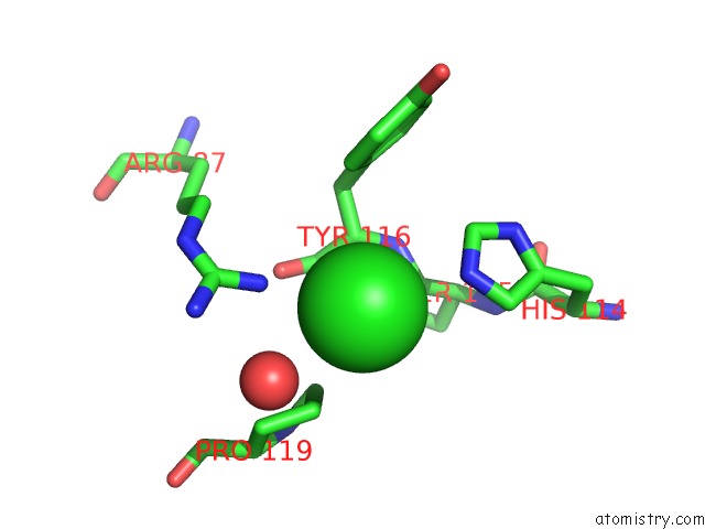
Mono view
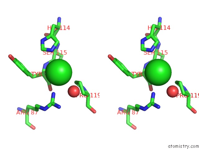
Stereo pair view
Mono view
Stereo pair view
A full contact list of Chlorine with other atoms in the Cl binding
site number 1 of Type II Dehydroquinase From Mycobacterium Tuberculosis Complexed with 2,3-Anhydro-Quinic Acid within 5.0Å range:
|
Chlorine binding site 2 out of 2 in 1h0r
Go back to
Chlorine binding site 2 out
of 2 in the Type II Dehydroquinase From Mycobacterium Tuberculosis Complexed with 2,3-Anhydro-Quinic Acid
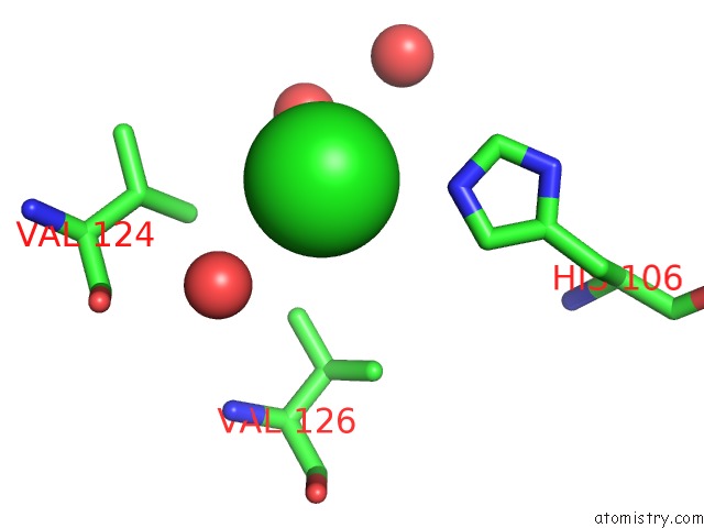
Mono view
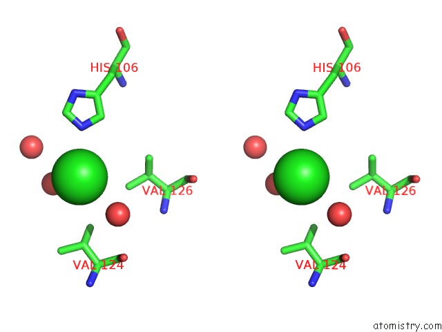
Stereo pair view
Mono view
Stereo pair view
A full contact list of Chlorine with other atoms in the Cl binding
site number 2 of Type II Dehydroquinase From Mycobacterium Tuberculosis Complexed with 2,3-Anhydro-Quinic Acid within 5.0Å range:
|
Reference:
D.A.Robinson,
A.W.Roszak,
M.Frederickson,
C.Abell,
J.R.Coggins,
A.J.Lapthorn.
Structural Basis For Selectivity of Oxime Based Inhibitors Towards Type II Dehydroquinase From Mycobacterium Tuberculosis To Be Published.
Page generated: Thu Jul 10 17:10:04 2025
Last articles
Fe in 7EMTFe in 7E5Z
Fe in 7EK4
Fe in 7EK5
Fe in 7EF8
Fe in 7EF9
Fe in 7EHX
Fe in 7EGN
Fe in 7EEH
Fe in 7EDA