Chlorine »
PDB 1go6-1hav »
1h3m »
Chlorine in PDB 1h3m: Structure of 4-Diphosphocytidyl-2C-Methyl-D-Erythritol Synthetase
Enzymatic activity of Structure of 4-Diphosphocytidyl-2C-Methyl-D-Erythritol Synthetase
All present enzymatic activity of Structure of 4-Diphosphocytidyl-2C-Methyl-D-Erythritol Synthetase:
2.7.7.60;
2.7.7.60;
Protein crystallography data
The structure of Structure of 4-Diphosphocytidyl-2C-Methyl-D-Erythritol Synthetase, PDB code: 1h3m
was solved by
L.E.Kemp,
C.S.Bond,
W.N.Hunter,
with X-Ray Crystallography technique. A brief refinement statistics is given in the table below:
| Resolution Low / High (Å) | 20.00 / 2.4 |
| Space group | P 41 21 2 |
| Cell size a, b, c (Å), α, β, γ (°) | 73.604, 73.604, 175.562, 90.00, 90.00, 90.00 |
| R / Rfree (%) | 23.7 / 33.3 |
Chlorine Binding Sites:
The binding sites of Chlorine atom in the Structure of 4-Diphosphocytidyl-2C-Methyl-D-Erythritol Synthetase
(pdb code 1h3m). This binding sites where shown within
5.0 Angstroms radius around Chlorine atom.
In total 5 binding sites of Chlorine where determined in the Structure of 4-Diphosphocytidyl-2C-Methyl-D-Erythritol Synthetase, PDB code: 1h3m:
Jump to Chlorine binding site number: 1; 2; 3; 4; 5;
In total 5 binding sites of Chlorine where determined in the Structure of 4-Diphosphocytidyl-2C-Methyl-D-Erythritol Synthetase, PDB code: 1h3m:
Jump to Chlorine binding site number: 1; 2; 3; 4; 5;
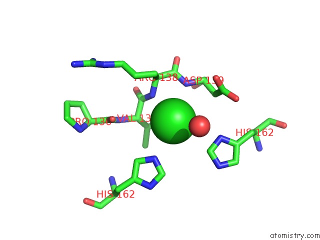
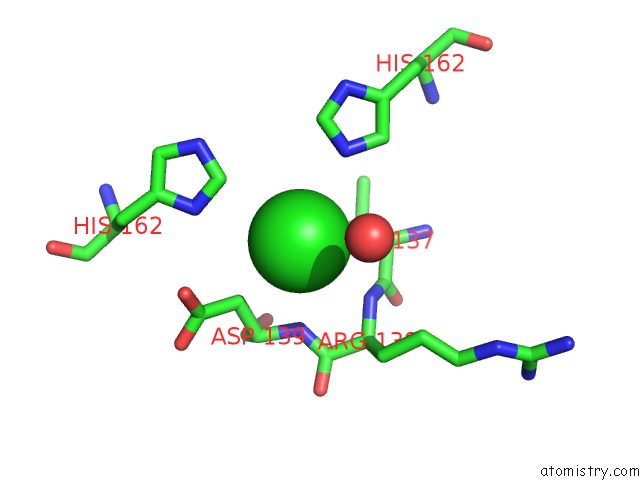
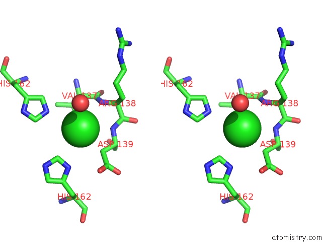
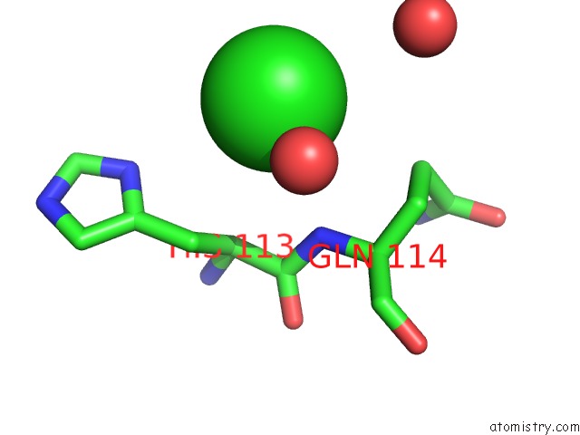
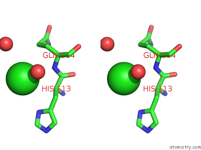
Chlorine binding site 1 out of 5 in 1h3m
Go back to
Chlorine binding site 1 out
of 5 in the Structure of 4-Diphosphocytidyl-2C-Methyl-D-Erythritol Synthetase
Mono view
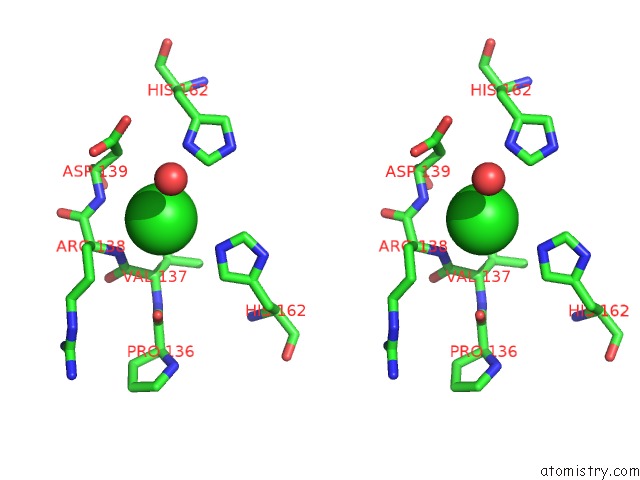
Stereo pair view
Mono view
Stereo pair view
A full contact list of Chlorine with other atoms in the Cl binding
site number 1 of Structure of 4-Diphosphocytidyl-2C-Methyl-D-Erythritol Synthetase within 5.0Å range:
|
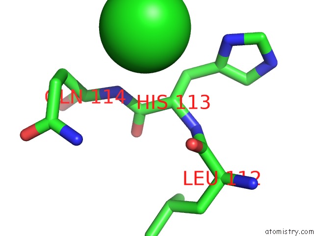
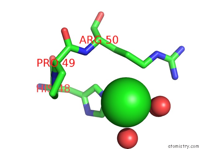
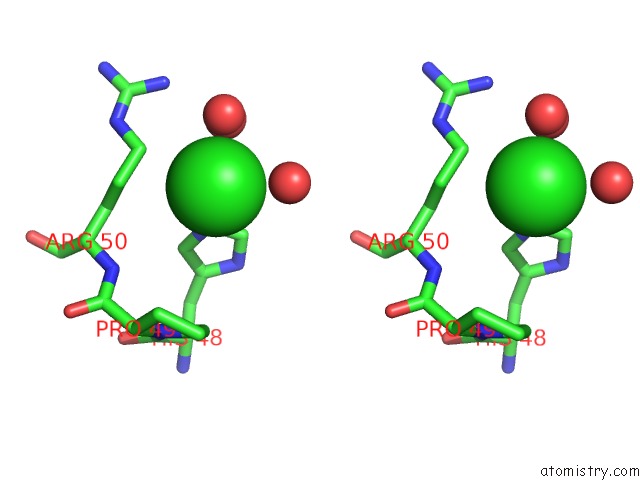
Chlorine binding site 2 out of 5 in 1h3m
Go back to
Chlorine binding site 2 out
of 5 in the Structure of 4-Diphosphocytidyl-2C-Methyl-D-Erythritol Synthetase
Mono view
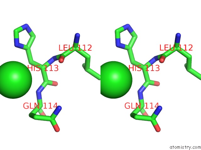
Stereo pair view
Mono view
Stereo pair view
A full contact list of Chlorine with other atoms in the Cl binding
site number 2 of Structure of 4-Diphosphocytidyl-2C-Methyl-D-Erythritol Synthetase within 5.0Å range:
|
Chlorine binding site 3 out of 5 in 1h3m
Go back to
Chlorine binding site 3 out
of 5 in the Structure of 4-Diphosphocytidyl-2C-Methyl-D-Erythritol Synthetase
Mono view
Stereo pair view
Mono view
Stereo pair view
A full contact list of Chlorine with other atoms in the Cl binding
site number 3 of Structure of 4-Diphosphocytidyl-2C-Methyl-D-Erythritol Synthetase within 5.0Å range:
|
Chlorine binding site 4 out of 5 in 1h3m
Go back to
Chlorine binding site 4 out
of 5 in the Structure of 4-Diphosphocytidyl-2C-Methyl-D-Erythritol Synthetase
Mono view
Stereo pair view
Mono view
Stereo pair view
A full contact list of Chlorine with other atoms in the Cl binding
site number 4 of Structure of 4-Diphosphocytidyl-2C-Methyl-D-Erythritol Synthetase within 5.0Å range:
|
Chlorine binding site 5 out of 5 in 1h3m
Go back to
Chlorine binding site 5 out
of 5 in the Structure of 4-Diphosphocytidyl-2C-Methyl-D-Erythritol Synthetase
Mono view
Stereo pair view
Mono view
Stereo pair view
A full contact list of Chlorine with other atoms in the Cl binding
site number 5 of Structure of 4-Diphosphocytidyl-2C-Methyl-D-Erythritol Synthetase within 5.0Å range:
|
Reference:
L.E.Kemp,
C.S.Bond,
W.N.Hunter.
Structure of A Tetragonal Crystal Form of Escherichia Coli 2-C-Methyl-D-Erythritol 4- Phosphate Cytidylyltransferase Acta Crystallogr.,Sect.D V. 59 607 2003.
ISSN: ISSN 0907-4449
PubMed: 12595740
DOI: 10.1107/S090744490202365X
Page generated: Thu Jul 10 17:10:45 2025
ISSN: ISSN 0907-4449
PubMed: 12595740
DOI: 10.1107/S090744490202365X
Last articles
Fe in 7F3WFe in 7F3H
Fe in 7F6X
Fe in 7EW6
Fe in 7EWK
Fe in 7F2Z
Fe in 7F30
Fe in 7F2Y
Fe in 7EYW
Fe in 7F0L