Chlorine »
PDB 1hba-1i3k »
1hxn »
Chlorine in PDB 1hxn: 1.8 Angstroms Crystal Structure of the C-Terminal Domain of Rabbit Serum Hemopexin
Protein crystallography data
The structure of 1.8 Angstroms Crystal Structure of the C-Terminal Domain of Rabbit Serum Hemopexin, PDB code: 1hxn
was solved by
H.R.Faber,
E.N.Baker,
with X-Ray Crystallography technique. A brief refinement statistics is given in the table below:
| Resolution Low / High (Å) | 20.00 / 1.80 |
| Space group | P 21 21 21 |
| Cell size a, b, c (Å), α, β, γ (°) | 43.600, 62.800, 80.600, 90.00, 90.00, 90.00 |
| R / Rfree (%) | n/a / n/a |
Other elements in 1hxn:
The structure of 1.8 Angstroms Crystal Structure of the C-Terminal Domain of Rabbit Serum Hemopexin also contains other interesting chemical elements:
| Sodium | (Na) | 2 atoms |
Chlorine Binding Sites:
The binding sites of Chlorine atom in the 1.8 Angstroms Crystal Structure of the C-Terminal Domain of Rabbit Serum Hemopexin
(pdb code 1hxn). This binding sites where shown within
5.0 Angstroms radius around Chlorine atom.
In total only one binding site of Chlorine was determined in the 1.8 Angstroms Crystal Structure of the C-Terminal Domain of Rabbit Serum Hemopexin, PDB code: 1hxn:
In total only one binding site of Chlorine was determined in the 1.8 Angstroms Crystal Structure of the C-Terminal Domain of Rabbit Serum Hemopexin, PDB code: 1hxn:
Chlorine binding site 1 out of 1 in 1hxn
Go back to
Chlorine binding site 1 out
of 1 in the 1.8 Angstroms Crystal Structure of the C-Terminal Domain of Rabbit Serum Hemopexin
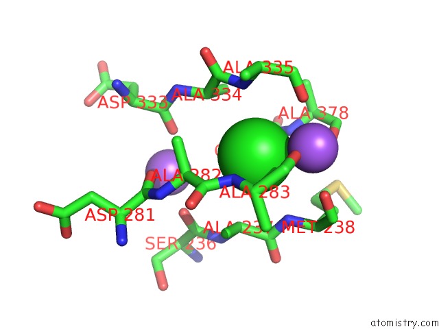
Mono view
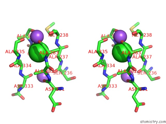
Stereo pair view
Mono view
Stereo pair view
A full contact list of Chlorine with other atoms in the Cl binding
site number 1 of 1.8 Angstroms Crystal Structure of the C-Terminal Domain of Rabbit Serum Hemopexin within 5.0Å range:
|
Reference:
H.R.Faber,
C.R.Groom,
H.M.Baker,
W.T.Morgan,
A.Smith,
E.N.Baker.
1.8 A Crystal Structure of the C-Terminal Domain of Rabbit Serum Haemopexin. Structure V. 3 551 1995.
ISSN: ISSN 0969-2126
PubMed: 8590016
DOI: 10.1016/S0969-2126(01)00189-7
Page generated: Thu Jul 10 17:18:01 2025
ISSN: ISSN 0969-2126
PubMed: 8590016
DOI: 10.1016/S0969-2126(01)00189-7
Last articles
Fe in 6ZKLFe in 6ZKI
Fe in 6ZKK
Fe in 6ZKJ
Fe in 6ZKH
Fe in 6ZKG
Fe in 6ZKF
Fe in 6ZKE
Fe in 6ZKD
Fe in 6ZKC