Chlorine »
PDB 1i3l-1iqi »
1i7d »
Chlorine in PDB 1i7d: Noncovalent Complex of E.Coli Dna Topoisomerase III with An 8-Base Single-Stranded Dna Oligonucleotide
Enzymatic activity of Noncovalent Complex of E.Coli Dna Topoisomerase III with An 8-Base Single-Stranded Dna Oligonucleotide
All present enzymatic activity of Noncovalent Complex of E.Coli Dna Topoisomerase III with An 8-Base Single-Stranded Dna Oligonucleotide:
5.99.1.2;
5.99.1.2;
Protein crystallography data
The structure of Noncovalent Complex of E.Coli Dna Topoisomerase III with An 8-Base Single-Stranded Dna Oligonucleotide, PDB code: 1i7d
was solved by
A.Changela,
R.J.Digate,
A.Mondragon,
with X-Ray Crystallography technique. A brief refinement statistics is given in the table below:
| Resolution Low / High (Å) | 29.63 / 2.05 |
| Space group | C 1 2 1 |
| Cell size a, b, c (Å), α, β, γ (°) | 122.011, 60.831, 125.358, 90.00, 90.73, 90.00 |
| R / Rfree (%) | 23.3 / 26 |
Chlorine Binding Sites:
The binding sites of Chlorine atom in the Noncovalent Complex of E.Coli Dna Topoisomerase III with An 8-Base Single-Stranded Dna Oligonucleotide
(pdb code 1i7d). This binding sites where shown within
5.0 Angstroms radius around Chlorine atom.
In total only one binding site of Chlorine was determined in the Noncovalent Complex of E.Coli Dna Topoisomerase III with An 8-Base Single-Stranded Dna Oligonucleotide, PDB code: 1i7d:
In total only one binding site of Chlorine was determined in the Noncovalent Complex of E.Coli Dna Topoisomerase III with An 8-Base Single-Stranded Dna Oligonucleotide, PDB code: 1i7d:
Chlorine binding site 1 out of 1 in 1i7d
Go back to
Chlorine binding site 1 out
of 1 in the Noncovalent Complex of E.Coli Dna Topoisomerase III with An 8-Base Single-Stranded Dna Oligonucleotide
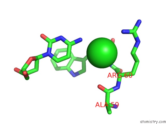
Mono view
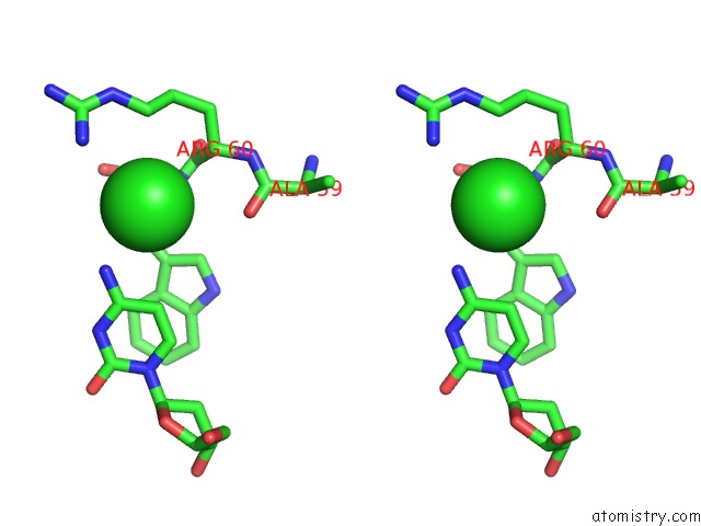
Stereo pair view
Mono view
Stereo pair view
A full contact list of Chlorine with other atoms in the Cl binding
site number 1 of Noncovalent Complex of E.Coli Dna Topoisomerase III with An 8-Base Single-Stranded Dna Oligonucleotide within 5.0Å range:
|
Reference:
A.Changela,
R.J.Digate,
A.Mondragon.
Crystal Structure of A Complex of A Type Ia Dna Topoisomerase with A Single-Stranded Dna Molecule. Nature V. 411 1077 2001.
ISSN: ISSN 0028-0836
PubMed: 11429611
DOI: 10.1038/35082615
Page generated: Thu Jul 10 17:21:35 2025
ISSN: ISSN 0028-0836
PubMed: 11429611
DOI: 10.1038/35082615
Last articles
K in 9NESK in 9PHG
K in 9NEI
K in 9NED
K in 9NEC
K in 9NEG
K in 9CWU
K in 9CVB
K in 9CVA
K in 9COM