Chlorine »
PDB 1k8a-1kv1 »
1ko1 »
Chlorine in PDB 1ko1: Crystal Structure of Gluconate Kinase
Enzymatic activity of Crystal Structure of Gluconate Kinase
All present enzymatic activity of Crystal Structure of Gluconate Kinase:
2.7.1.12;
2.7.1.12;
Protein crystallography data
The structure of Crystal Structure of Gluconate Kinase, PDB code: 1ko1
was solved by
L.Kraft,
G.A.Sprenger,
Y.Lindqvist,
with X-Ray Crystallography technique. A brief refinement statistics is given in the table below:
| Resolution Low / High (Å) | 25.00 / 2.09 |
| Space group | C 1 2 1 |
| Cell size a, b, c (Å), α, β, γ (°) | 74.556, 79.167, 70.091, 90.00, 105.20, 90.00 |
| R / Rfree (%) | 21.8 / 28 |
Chlorine Binding Sites:
The binding sites of Chlorine atom in the Crystal Structure of Gluconate Kinase
(pdb code 1ko1). This binding sites where shown within
5.0 Angstroms radius around Chlorine atom.
In total 2 binding sites of Chlorine where determined in the Crystal Structure of Gluconate Kinase, PDB code: 1ko1:
Jump to Chlorine binding site number: 1; 2;
In total 2 binding sites of Chlorine where determined in the Crystal Structure of Gluconate Kinase, PDB code: 1ko1:
Jump to Chlorine binding site number: 1; 2;
Chlorine binding site 1 out of 2 in 1ko1
Go back to
Chlorine binding site 1 out
of 2 in the Crystal Structure of Gluconate Kinase
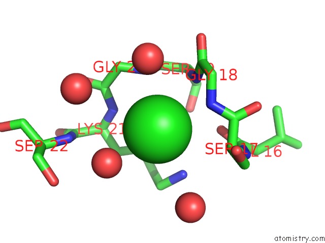
Mono view
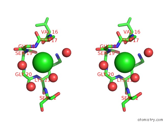
Stereo pair view
Mono view
Stereo pair view
A full contact list of Chlorine with other atoms in the Cl binding
site number 1 of Crystal Structure of Gluconate Kinase within 5.0Å range:
|
Chlorine binding site 2 out of 2 in 1ko1
Go back to
Chlorine binding site 2 out
of 2 in the Crystal Structure of Gluconate Kinase
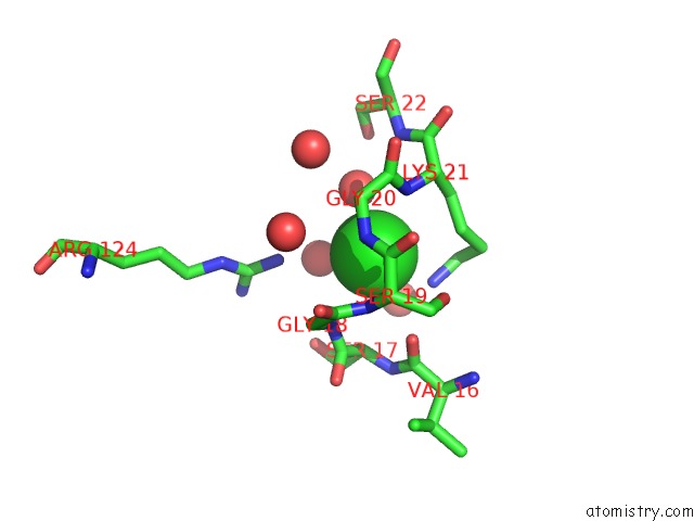
Mono view
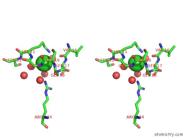
Stereo pair view
Mono view
Stereo pair view
A full contact list of Chlorine with other atoms in the Cl binding
site number 2 of Crystal Structure of Gluconate Kinase within 5.0Å range:
|
Reference:
L.Kraft,
G.A.Sprenger,
Y.Lindqvist.
Conformational Changes During the Catalytic Cycle of Gluconate Kinase As Revealed By X-Ray Crystallography. J.Mol.Biol. V. 318 1057 2002.
ISSN: ISSN 0022-2836
PubMed: 12054802
DOI: 10.1016/S0022-2836(02)00215-2
Page generated: Thu Jul 10 17:48:41 2025
ISSN: ISSN 0022-2836
PubMed: 12054802
DOI: 10.1016/S0022-2836(02)00215-2
Last articles
Mg in 5WPMMg in 5WPL
Mg in 5WNS
Mg in 5WNQ
Mg in 5WNR
Mg in 5WNP
Mg in 5WMB
Mg in 5WM8
Mg in 5WNO
Mg in 5WNI