Chlorine »
PDB 1mkp-1nc4 »
1mx5 »
Chlorine in PDB 1mx5: Crystal Structure of Human Liver Carboxylesterase in Complexed with Homatropine, A Cocaine Analogue
Enzymatic activity of Crystal Structure of Human Liver Carboxylesterase in Complexed with Homatropine, A Cocaine Analogue
All present enzymatic activity of Crystal Structure of Human Liver Carboxylesterase in Complexed with Homatropine, A Cocaine Analogue:
3.1.1.1;
3.1.1.1;
Protein crystallography data
The structure of Crystal Structure of Human Liver Carboxylesterase in Complexed with Homatropine, A Cocaine Analogue, PDB code: 1mx5
was solved by
S.Bencharit,
C.L.Morton,
Y.Xue,
P.M.Potter,
M.R.Redinbo,
with X-Ray Crystallography technique. A brief refinement statistics is given in the table below:
| Resolution Low / High (Å) | 19.96 / 2.80 |
| Space group | P 1 21 1 |
| Cell size a, b, c (Å), α, β, γ (°) | 55.400, 178.800, 199.600, 90.00, 90.20, 90.00 |
| R / Rfree (%) | 15.8 / 22.1 |
Chlorine Binding Sites:
The binding sites of Chlorine atom in the Crystal Structure of Human Liver Carboxylesterase in Complexed with Homatropine, A Cocaine Analogue
(pdb code 1mx5). This binding sites where shown within
5.0 Angstroms radius around Chlorine atom.
In total 2 binding sites of Chlorine where determined in the Crystal Structure of Human Liver Carboxylesterase in Complexed with Homatropine, A Cocaine Analogue, PDB code: 1mx5:
Jump to Chlorine binding site number: 1; 2;
In total 2 binding sites of Chlorine where determined in the Crystal Structure of Human Liver Carboxylesterase in Complexed with Homatropine, A Cocaine Analogue, PDB code: 1mx5:
Jump to Chlorine binding site number: 1; 2;
Chlorine binding site 1 out of 2 in 1mx5
Go back to
Chlorine binding site 1 out
of 2 in the Crystal Structure of Human Liver Carboxylesterase in Complexed with Homatropine, A Cocaine Analogue
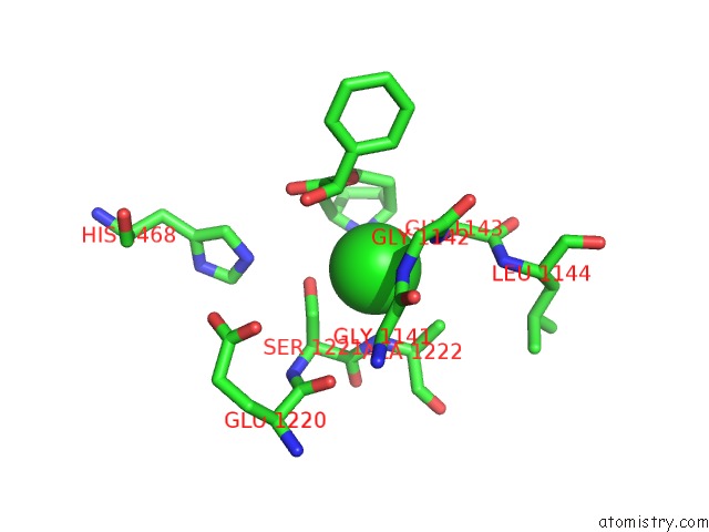
Mono view
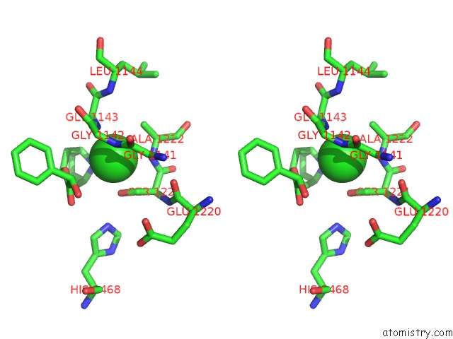
Stereo pair view
Mono view
Stereo pair view
A full contact list of Chlorine with other atoms in the Cl binding
site number 1 of Crystal Structure of Human Liver Carboxylesterase in Complexed with Homatropine, A Cocaine Analogue within 5.0Å range:
|
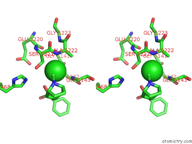
Chlorine binding site 2 out of 2 in 1mx5
Go back to
Chlorine binding site 2 out
of 2 in the Crystal Structure of Human Liver Carboxylesterase in Complexed with Homatropine, A Cocaine Analogue
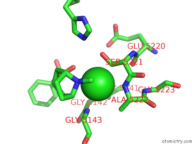
Mono view
Stereo pair view
Mono view
Stereo pair view
A full contact list of Chlorine with other atoms in the Cl binding
site number 2 of Crystal Structure of Human Liver Carboxylesterase in Complexed with Homatropine, A Cocaine Analogue within 5.0Å range:
|
Reference:
S.Bencharit,
C.L.Morton,
Y.Xue,
P.M.Potter,
M.R.Redinbo.
Structural Basis of Heroin and Cocaine Metabolism By A Promiscuous Human Drug-Processing Enzyme Nat.Struct.Biol. V. 10 349 2003.
ISSN: ISSN 1072-8368
PubMed: 12679808
DOI: 10.1038/NSB919
Page generated: Thu Jul 10 18:17:34 2025
ISSN: ISSN 1072-8368
PubMed: 12679808
DOI: 10.1038/NSB919
Last articles
K in 7FS0K in 7FRZ
K in 7FRY
K in 7FRX
K in 7FRW
K in 7FRV
K in 7F8Z
K in 7FCV
K in 7F7Y
K in 7FHA