Chlorine »
PDB 1pu6-1qhu »
1pu6 »
Chlorine in PDB 1pu6: Crystal Structure of H.Pylori 3-Methyladenine Dna Glycosylase (Magiii)
Protein crystallography data
The structure of Crystal Structure of H.Pylori 3-Methyladenine Dna Glycosylase (Magiii), PDB code: 1pu6
was solved by
B.F.Eichman,
E.J.O'rourke,
J.P.Radicella,
T.Ellenberger,
with X-Ray Crystallography technique. A brief refinement statistics is given in the table below:
| Resolution Low / High (Å) | 24.41 / 1.64 |
| Space group | C 1 2 1 |
| Cell size a, b, c (Å), α, β, γ (°) | 146.605, 44.399, 81.516, 90.00, 106.40, 90.00 |
| R / Rfree (%) | 15.1 / 18.5 |
Chlorine Binding Sites:
The binding sites of Chlorine atom in the Crystal Structure of H.Pylori 3-Methyladenine Dna Glycosylase (Magiii)
(pdb code 1pu6). This binding sites where shown within
5.0 Angstroms radius around Chlorine atom.
In total only one binding site of Chlorine was determined in the Crystal Structure of H.Pylori 3-Methyladenine Dna Glycosylase (Magiii), PDB code: 1pu6:
In total only one binding site of Chlorine was determined in the Crystal Structure of H.Pylori 3-Methyladenine Dna Glycosylase (Magiii), PDB code: 1pu6:
Chlorine binding site 1 out of 1 in 1pu6
Go back to
Chlorine binding site 1 out
of 1 in the Crystal Structure of H.Pylori 3-Methyladenine Dna Glycosylase (Magiii)
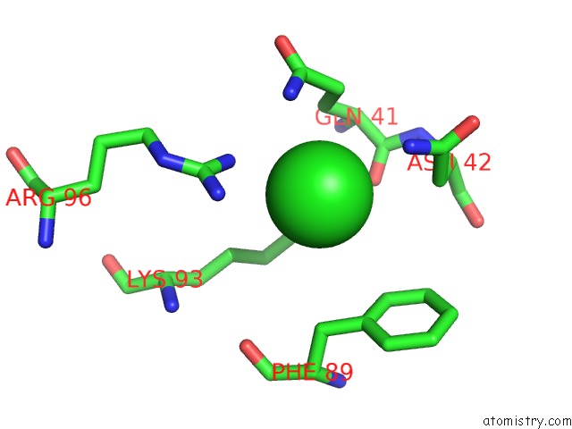
Mono view
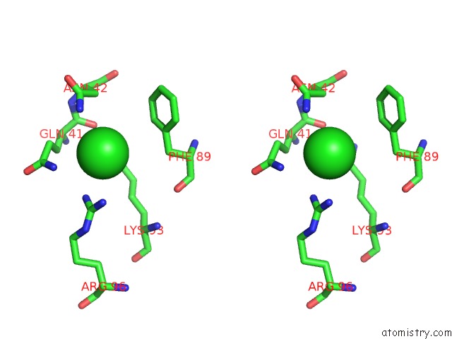
Stereo pair view
Mono view
Stereo pair view
A full contact list of Chlorine with other atoms in the Cl binding
site number 1 of Crystal Structure of H.Pylori 3-Methyladenine Dna Glycosylase (Magiii) within 5.0Å range:
|
Reference:
B.F.Eichman,
E.J.O'rourke,
J.P.Radicella,
T.Ellenberger.
Crystal Structures of 3-Methyladenine Dna Glycosylase Magiii and the Recognition of Alkylated Bases Embo J. V. 22 4898 2003.
ISSN: ISSN 0261-4189
PubMed: 14517230
DOI: 10.1093/EMBOJ/CDG505
Page generated: Thu Jul 10 18:53:55 2025
ISSN: ISSN 0261-4189
PubMed: 14517230
DOI: 10.1093/EMBOJ/CDG505
Last articles
Fe in 2CKJFe in 2CTH
Fe in 2CXB
Fe in 2CW3
Fe in 2CW2
Fe in 2CKF
Fe in 2CSG
Fe in 2CPP
Fe in 2CPO
Fe in 2CLB