Chlorine »
PDB 1pu6-1qhu »
1qgw »
Chlorine in PDB 1qgw: Crystal Structure of Phycoerythrin 545 From the Marine Cryptophyte Rhodomonas CS24
Protein crystallography data
The structure of Crystal Structure of Phycoerythrin 545 From the Marine Cryptophyte Rhodomonas CS24, PDB code: 1qgw
was solved by
S.J.Harrop,
K.E.Wilk,
R.G.Hiller,
P.M.G.Curmi,
with X-Ray Crystallography technique. A brief refinement statistics is given in the table below:
| Resolution Low / High (Å) | 20.00 / 1.63 |
| Space group | P 21 21 21 |
| Cell size a, b, c (Å), α, β, γ (°) | 63.030, 82.630, 89.550, 90.00, 90.00, 90.00 |
| R / Rfree (%) | 14.9 / 18.8 |
Other elements in 1qgw:
The structure of Crystal Structure of Phycoerythrin 545 From the Marine Cryptophyte Rhodomonas CS24 also contains other interesting chemical elements:
| Magnesium | (Mg) | 2 atoms |
Chlorine Binding Sites:
The binding sites of Chlorine atom in the Crystal Structure of Phycoerythrin 545 From the Marine Cryptophyte Rhodomonas CS24
(pdb code 1qgw). This binding sites where shown within
5.0 Angstroms radius around Chlorine atom.
In total only one binding site of Chlorine was determined in the Crystal Structure of Phycoerythrin 545 From the Marine Cryptophyte Rhodomonas CS24, PDB code: 1qgw:
In total only one binding site of Chlorine was determined in the Crystal Structure of Phycoerythrin 545 From the Marine Cryptophyte Rhodomonas CS24, PDB code: 1qgw:
Chlorine binding site 1 out of 1 in 1qgw
Go back to
Chlorine binding site 1 out
of 1 in the Crystal Structure of Phycoerythrin 545 From the Marine Cryptophyte Rhodomonas CS24
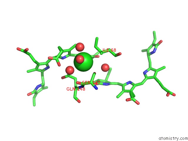
Mono view
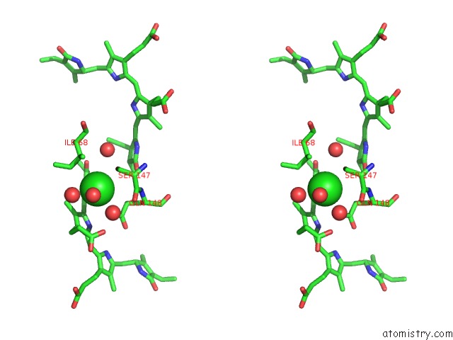
Stereo pair view
Mono view
Stereo pair view
A full contact list of Chlorine with other atoms in the Cl binding
site number 1 of Crystal Structure of Phycoerythrin 545 From the Marine Cryptophyte Rhodomonas CS24 within 5.0Å range:
|
Reference:
K.E.Wilk,
S.J.Harrop,
L.Jankova,
D.Edler,
G.Keenan,
F.Sharples,
R.G.Hiller,
P.M.Curmi.
Evolution of A Light-Harvesting Protein By Addition of New Subunits and Rearrangement of Conserved Elements: Crystal Structure of A Cryptophyte Phycoerythrin at 1.63-A Resolution. Proc.Natl.Acad.Sci.Usa V. 96 8901 1999.
ISSN: ISSN 0027-8424
PubMed: 10430868
DOI: 10.1073/PNAS.96.16.8901
Page generated: Thu Jul 10 19:02:36 2025
ISSN: ISSN 0027-8424
PubMed: 10430868
DOI: 10.1073/PNAS.96.16.8901
Last articles
Fe in 2YXOFe in 2YRS
Fe in 2YXC
Fe in 2YNM
Fe in 2YVJ
Fe in 2YP1
Fe in 2YU2
Fe in 2YU1
Fe in 2YQB
Fe in 2YOO