Chlorine »
PDB 1rky-1s8f »
1s4q »
Chlorine in PDB 1s4q: Crystal Structure of Guanylate Kinase From Mycobacterium Tuberculosis (RV1389)
Enzymatic activity of Crystal Structure of Guanylate Kinase From Mycobacterium Tuberculosis (RV1389)
All present enzymatic activity of Crystal Structure of Guanylate Kinase From Mycobacterium Tuberculosis (RV1389):
2.7.4.8;
2.7.4.8;
Protein crystallography data
The structure of Crystal Structure of Guanylate Kinase From Mycobacterium Tuberculosis (RV1389), PDB code: 1s4q
was solved by
S.Chan,
M.R.Sawaya,
L.J.Perry,
D.Eisenberg,
Tb Structural Genomicsconsortium (Tbsgc),
with X-Ray Crystallography technique. A brief refinement statistics is given in the table below:
| Resolution Low / High (Å) | 79.06 / 2.16 |
| Space group | I 2 3 |
| Cell size a, b, c (Å), α, β, γ (°) | 112.126, 112.126, 112.126, 90.00, 90.00, 90.00 |
| R / Rfree (%) | 17.7 / 23.1 |
Chlorine Binding Sites:
The binding sites of Chlorine atom in the Crystal Structure of Guanylate Kinase From Mycobacterium Tuberculosis (RV1389)
(pdb code 1s4q). This binding sites where shown within
5.0 Angstroms radius around Chlorine atom.
In total only one binding site of Chlorine was determined in the Crystal Structure of Guanylate Kinase From Mycobacterium Tuberculosis (RV1389), PDB code: 1s4q:
In total only one binding site of Chlorine was determined in the Crystal Structure of Guanylate Kinase From Mycobacterium Tuberculosis (RV1389), PDB code: 1s4q:
Chlorine binding site 1 out of 1 in 1s4q
Go back to
Chlorine binding site 1 out
of 1 in the Crystal Structure of Guanylate Kinase From Mycobacterium Tuberculosis (RV1389)
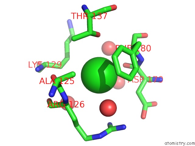
Mono view
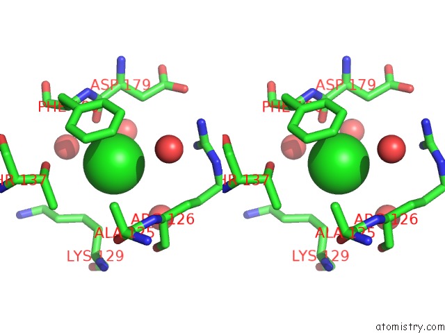
Stereo pair view
Mono view
Stereo pair view
A full contact list of Chlorine with other atoms in the Cl binding
site number 1 of Crystal Structure of Guanylate Kinase From Mycobacterium Tuberculosis (RV1389) within 5.0Å range:
|
Reference:
S.Chan,
M.R.Sawaya,
L.J.Perry,
D.Eisenberg.
Crystal Structure of Guanylate Kinase From Mycobacterium Tuberculosis To Be Published.
Page generated: Thu Jul 10 19:20:18 2025
Last articles
Fe in 8DZLFe in 8DWF
Fe in 8DZK
Fe in 8DY9
Fe in 8DY7
Fe in 8DYC
Fe in 8DYB
Fe in 8DWD
Fe in 8DWE
Fe in 8DWR