Chlorine »
PDB 1swz-1tgx »
1tgx »
Chlorine in PDB 1tgx: X-Ray Structure at 1.55 A of Toxin Gamma, A Cardiotoxin From Naja Nigricollis Venom. Crystal Packing Reveals A Model For Insertion Into Membranes
Protein crystallography data
The structure of X-Ray Structure at 1.55 A of Toxin Gamma, A Cardiotoxin From Naja Nigricollis Venom. Crystal Packing Reveals A Model For Insertion Into Membranes, PDB code: 1tgx
was solved by
A.Bilwes,
B.Rees,
D.Moras,
with X-Ray Crystallography technique. A brief refinement statistics is given in the table below:
| Resolution Low / High (Å) | 5.00 / 1.55 |
| Space group | C 1 2 1 |
| Cell size a, b, c (Å), α, β, γ (°) | 78.670, 40.710, 56.020, 90.00, 117.08, 90.00 |
| R / Rfree (%) | n/a / n/a |
Chlorine Binding Sites:
Pages:
>>> Page 1 <<< Page 2, Binding sites: 11 - 14;Binding sites:
The binding sites of Chlorine atom in the X-Ray Structure at 1.55 A of Toxin Gamma, A Cardiotoxin From Naja Nigricollis Venom. Crystal Packing Reveals A Model For Insertion Into Membranes (pdb code 1tgx). This binding sites where shown within 5.0 Angstroms radius around Chlorine atom.In total 14 binding sites of Chlorine where determined in the X-Ray Structure at 1.55 A of Toxin Gamma, A Cardiotoxin From Naja Nigricollis Venom. Crystal Packing Reveals A Model For Insertion Into Membranes, PDB code: 1tgx:
Jump to Chlorine binding site number: 1; 2; 3; 4; 5; 6; 7; 8; 9; 10;
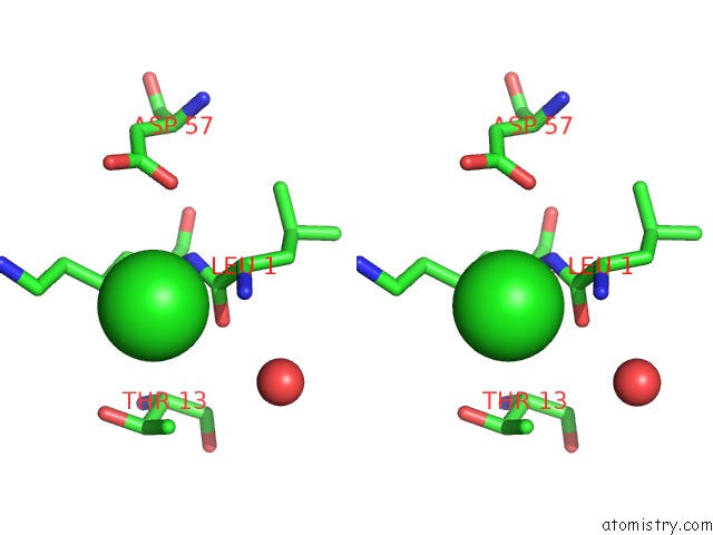
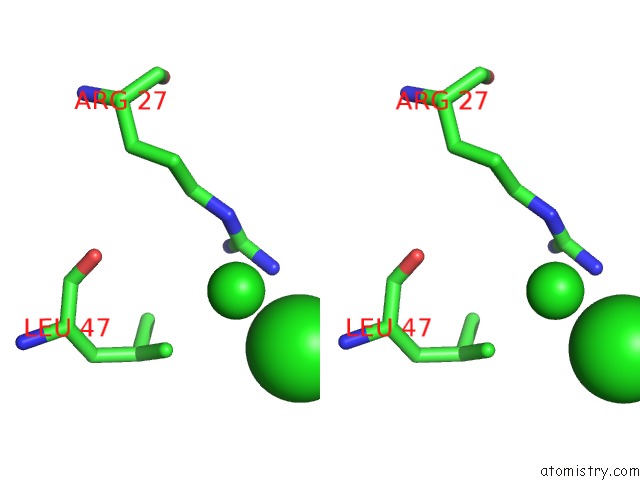
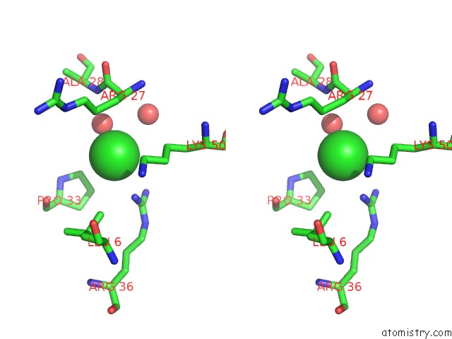
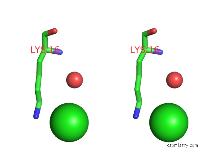
Chlorine binding site 1 out of 14 in 1tgx
Go back to
Chlorine binding site 1 out
of 14 in the X-Ray Structure at 1.55 A of Toxin Gamma, A Cardiotoxin From Naja Nigricollis Venom. Crystal Packing Reveals A Model For Insertion Into Membranes

Mono view

Stereo pair view
Mono view
Stereo pair view
A full contact list of Chlorine with other atoms in the Cl binding
site number 1 of X-Ray Structure at 1.55 A of Toxin Gamma, A Cardiotoxin From Naja Nigricollis Venom. Crystal Packing Reveals A Model For Insertion Into Membranes within 5.0Å range:
|
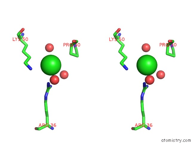
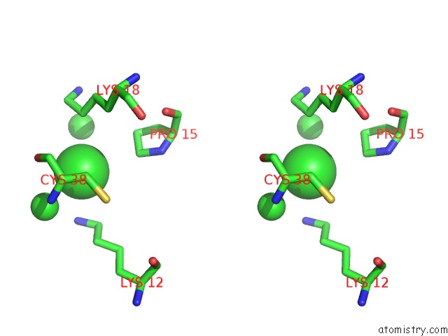
Chlorine binding site 2 out of 14 in 1tgx
Go back to
Chlorine binding site 2 out
of 14 in the X-Ray Structure at 1.55 A of Toxin Gamma, A Cardiotoxin From Naja Nigricollis Venom. Crystal Packing Reveals A Model For Insertion Into Membranes

Mono view
Stereo pair view
Mono view
Stereo pair view
A full contact list of Chlorine with other atoms in the Cl binding
site number 2 of X-Ray Structure at 1.55 A of Toxin Gamma, A Cardiotoxin From Naja Nigricollis Venom. Crystal Packing Reveals A Model For Insertion Into Membranes within 5.0Å range:
|
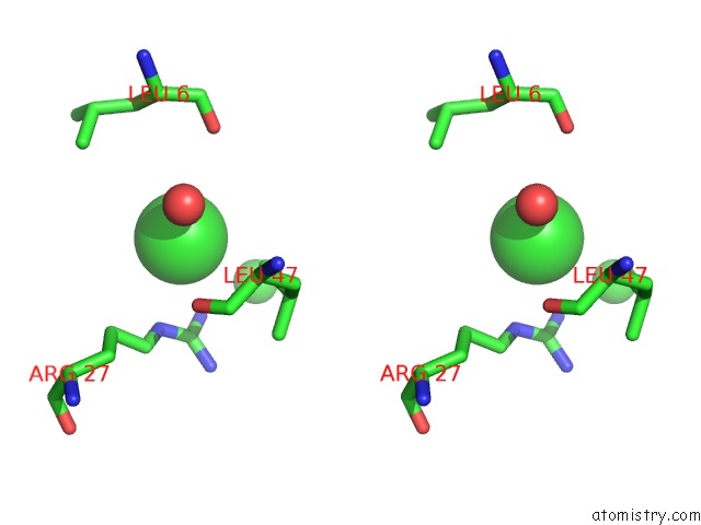
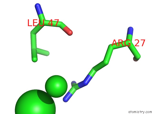
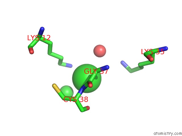
Chlorine binding site 3 out of 14 in 1tgx
Go back to
Chlorine binding site 3 out
of 14 in the X-Ray Structure at 1.55 A of Toxin Gamma, A Cardiotoxin From Naja Nigricollis Venom. Crystal Packing Reveals A Model For Insertion Into Membranes

Mono view
Stereo pair view
Mono view
Stereo pair view
A full contact list of Chlorine with other atoms in the Cl binding
site number 3 of X-Ray Structure at 1.55 A of Toxin Gamma, A Cardiotoxin From Naja Nigricollis Venom. Crystal Packing Reveals A Model For Insertion Into Membranes within 5.0Å range:
|
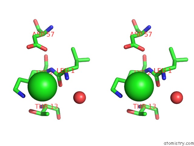
Chlorine binding site 4 out of 14 in 1tgx
Go back to
Chlorine binding site 4 out
of 14 in the X-Ray Structure at 1.55 A of Toxin Gamma, A Cardiotoxin From Naja Nigricollis Venom. Crystal Packing Reveals A Model For Insertion Into Membranes

Mono view
Stereo pair view
Mono view
Stereo pair view
A full contact list of Chlorine with other atoms in the Cl binding
site number 4 of X-Ray Structure at 1.55 A of Toxin Gamma, A Cardiotoxin From Naja Nigricollis Venom. Crystal Packing Reveals A Model For Insertion Into Membranes within 5.0Å range:
|
Chlorine binding site 5 out of 14 in 1tgx
Go back to
Chlorine binding site 5 out
of 14 in the X-Ray Structure at 1.55 A of Toxin Gamma, A Cardiotoxin From Naja Nigricollis Venom. Crystal Packing Reveals A Model For Insertion Into Membranes
Mono view
Stereo pair view
Mono view
Stereo pair view
A full contact list of Chlorine with other atoms in the Cl binding
site number 5 of X-Ray Structure at 1.55 A of Toxin Gamma, A Cardiotoxin From Naja Nigricollis Venom. Crystal Packing Reveals A Model For Insertion Into Membranes within 5.0Å range:
|
Chlorine binding site 6 out of 14 in 1tgx
Go back to
Chlorine binding site 6 out
of 14 in the X-Ray Structure at 1.55 A of Toxin Gamma, A Cardiotoxin From Naja Nigricollis Venom. Crystal Packing Reveals A Model For Insertion Into Membranes

Mono view
Stereo pair view
Mono view
Stereo pair view
A full contact list of Chlorine with other atoms in the Cl binding
site number 6 of X-Ray Structure at 1.55 A of Toxin Gamma, A Cardiotoxin From Naja Nigricollis Venom. Crystal Packing Reveals A Model For Insertion Into Membranes within 5.0Å range:
|
Chlorine binding site 7 out of 14 in 1tgx
Go back to
Chlorine binding site 7 out
of 14 in the X-Ray Structure at 1.55 A of Toxin Gamma, A Cardiotoxin From Naja Nigricollis Venom. Crystal Packing Reveals A Model For Insertion Into Membranes
Mono view

Stereo pair view
Mono view
Stereo pair view
A full contact list of Chlorine with other atoms in the Cl binding
site number 7 of X-Ray Structure at 1.55 A of Toxin Gamma, A Cardiotoxin From Naja Nigricollis Venom. Crystal Packing Reveals A Model For Insertion Into Membranes within 5.0Å range:
|
Chlorine binding site 8 out of 14 in 1tgx
Go back to
Chlorine binding site 8 out
of 14 in the X-Ray Structure at 1.55 A of Toxin Gamma, A Cardiotoxin From Naja Nigricollis Venom. Crystal Packing Reveals A Model For Insertion Into Membranes

Mono view
Stereo pair view
Mono view
Stereo pair view
A full contact list of Chlorine with other atoms in the Cl binding
site number 8 of X-Ray Structure at 1.55 A of Toxin Gamma, A Cardiotoxin From Naja Nigricollis Venom. Crystal Packing Reveals A Model For Insertion Into Membranes within 5.0Å range:
|
Chlorine binding site 9 out of 14 in 1tgx
Go back to
Chlorine binding site 9 out
of 14 in the X-Ray Structure at 1.55 A of Toxin Gamma, A Cardiotoxin From Naja Nigricollis Venom. Crystal Packing Reveals A Model For Insertion Into Membranes

Mono view
Stereo pair view
Mono view
Stereo pair view
A full contact list of Chlorine with other atoms in the Cl binding
site number 9 of X-Ray Structure at 1.55 A of Toxin Gamma, A Cardiotoxin From Naja Nigricollis Venom. Crystal Packing Reveals A Model For Insertion Into Membranes within 5.0Å range:
|
Chlorine binding site 10 out of 14 in 1tgx
Go back to
Chlorine binding site 10 out
of 14 in the X-Ray Structure at 1.55 A of Toxin Gamma, A Cardiotoxin From Naja Nigricollis Venom. Crystal Packing Reveals A Model For Insertion Into Membranes

Mono view
Stereo pair view
Mono view
Stereo pair view
A full contact list of Chlorine with other atoms in the Cl binding
site number 10 of X-Ray Structure at 1.55 A of Toxin Gamma, A Cardiotoxin From Naja Nigricollis Venom. Crystal Packing Reveals A Model For Insertion Into Membranes within 5.0Å range:
|
Reference:
A.Bilwes,
B.Rees,
D.Moras,
R.Menez,
A.Menez.
X-Ray Structure at 1.55 A of Toxin Gamma, A Cardiotoxin From Naja Nigricollis Venom. Crystal Packing Reveals A Model For Insertion Into Membranes. J.Mol.Biol. V. 239 122 1994.
ISSN: ISSN 0022-2836
PubMed: 8196041
DOI: 10.1006/JMBI.1994.1357
Page generated: Thu Jul 10 19:35:09 2025
ISSN: ISSN 0022-2836
PubMed: 8196041
DOI: 10.1006/JMBI.1994.1357
Last articles
Mg in 1LJ0Mg in 1LJX
Mg in 1LIJ
Mg in 1LII
Mg in 1LH0
Mg in 1LFD
Mg in 1L9J
Mg in 1LF5
Mg in 1LF0
Mg in 1LDF