Chlorine »
PDB 1ti7-1u33 »
1tpk »
Chlorine in PDB 1tpk: Crystal Structure of the Kringle-2 Domain of Tissue Plasminogen Activator at 2.4-Angstroms Resolution
Enzymatic activity of Crystal Structure of the Kringle-2 Domain of Tissue Plasminogen Activator at 2.4-Angstroms Resolution
All present enzymatic activity of Crystal Structure of the Kringle-2 Domain of Tissue Plasminogen Activator at 2.4-Angstroms Resolution:
3.4.21.31;
3.4.21.31;
Protein crystallography data
The structure of Crystal Structure of the Kringle-2 Domain of Tissue Plasminogen Activator at 2.4-Angstroms Resolution, PDB code: 1tpk
was solved by
A.M.De Vos,
M.H.Ultsch,
R.F.Kelley,
K.Padmanabhan,
A.Tulinsky,
M.L.Westbrook,
A.A.Kossiakoff,
with X-Ray Crystallography technique. A brief refinement statistics is given in the table below:
| Resolution Low / High (Å) | 10.00 / 2.40 |
| Space group | P 1 21 1 |
| Cell size a, b, c (Å), α, β, γ (°) | 54.800, 63.580, 46.580, 90.00, 106.73, 90.00 |
| R / Rfree (%) | 18.4 / n/a |
Chlorine Binding Sites:
The binding sites of Chlorine atom in the Crystal Structure of the Kringle-2 Domain of Tissue Plasminogen Activator at 2.4-Angstroms Resolution
(pdb code 1tpk). This binding sites where shown within
5.0 Angstroms radius around Chlorine atom.
In total 3 binding sites of Chlorine where determined in the Crystal Structure of the Kringle-2 Domain of Tissue Plasminogen Activator at 2.4-Angstroms Resolution, PDB code: 1tpk:
Jump to Chlorine binding site number: 1; 2; 3;
In total 3 binding sites of Chlorine where determined in the Crystal Structure of the Kringle-2 Domain of Tissue Plasminogen Activator at 2.4-Angstroms Resolution, PDB code: 1tpk:
Jump to Chlorine binding site number: 1; 2; 3;
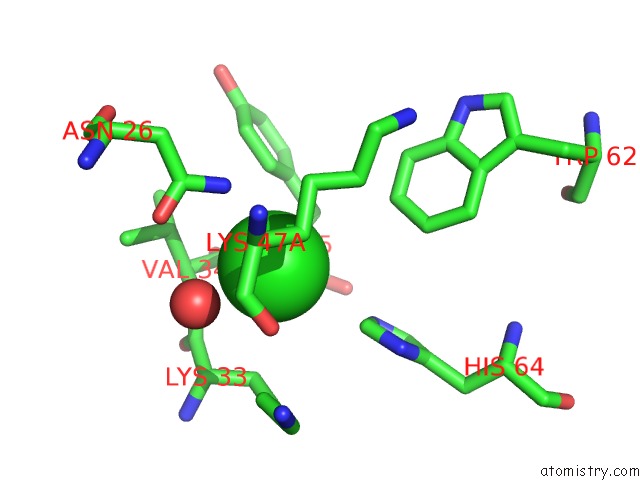
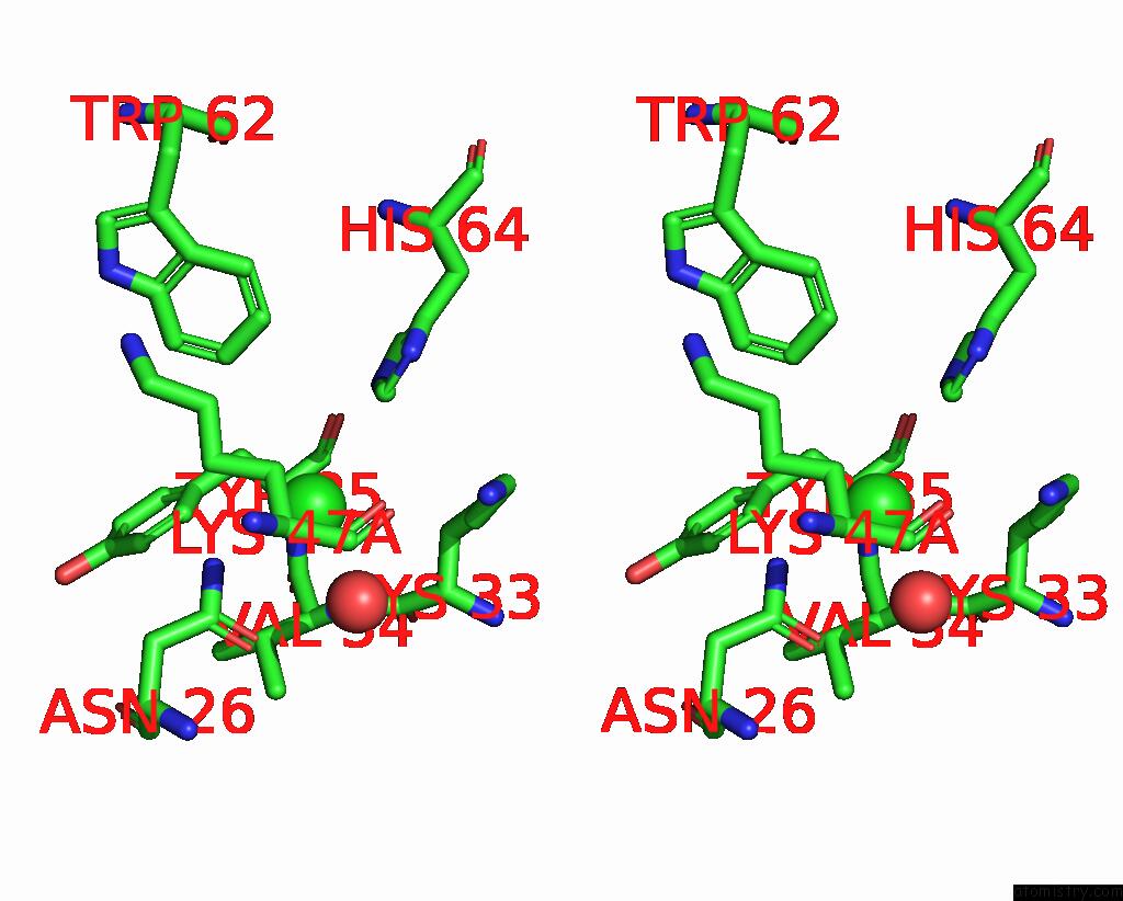
Chlorine binding site 1 out of 3 in 1tpk
Go back to
Chlorine binding site 1 out
of 3 in the Crystal Structure of the Kringle-2 Domain of Tissue Plasminogen Activator at 2.4-Angstroms Resolution
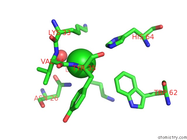
Mono view
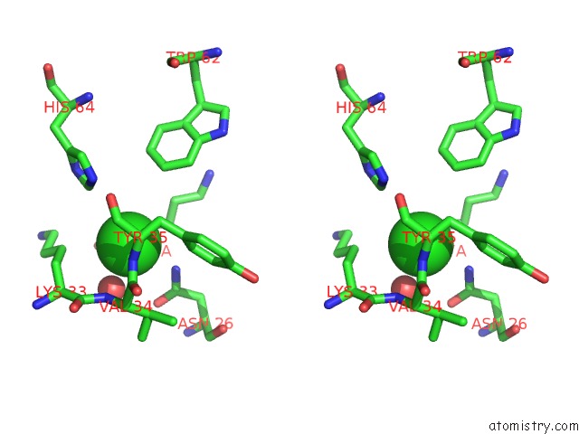
Stereo pair view
Mono view
Stereo pair view
A full contact list of Chlorine with other atoms in the Cl binding
site number 1 of Crystal Structure of the Kringle-2 Domain of Tissue Plasminogen Activator at 2.4-Angstroms Resolution within 5.0Å range:
|
Chlorine binding site 2 out of 3 in 1tpk
Go back to
Chlorine binding site 2 out
of 3 in the Crystal Structure of the Kringle-2 Domain of Tissue Plasminogen Activator at 2.4-Angstroms Resolution
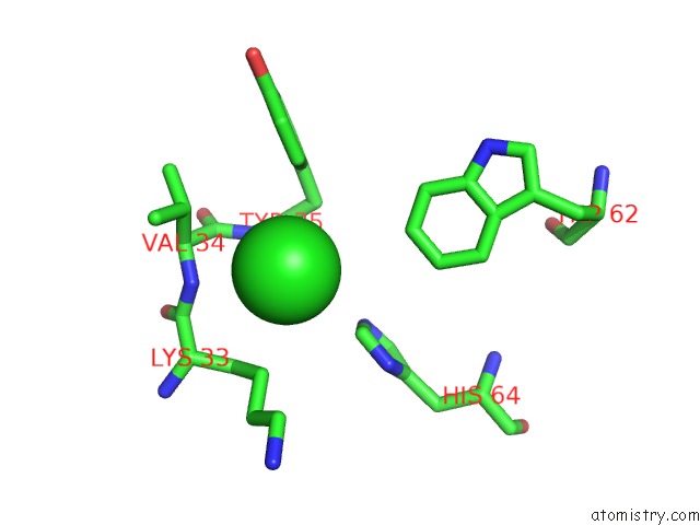
Mono view
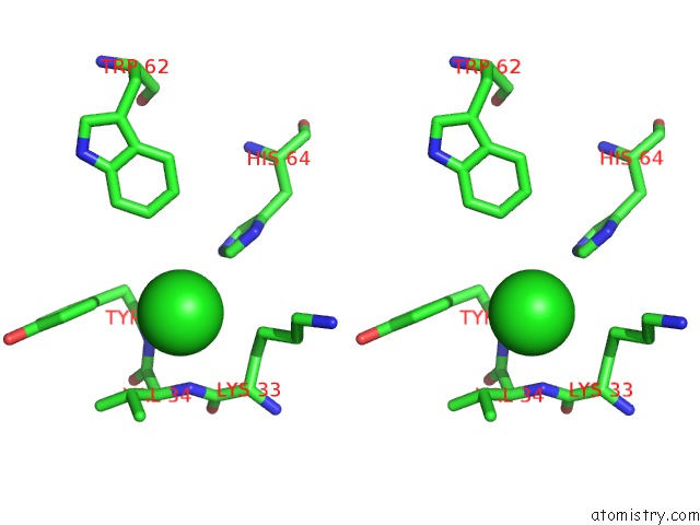
Stereo pair view
Mono view
Stereo pair view
A full contact list of Chlorine with other atoms in the Cl binding
site number 2 of Crystal Structure of the Kringle-2 Domain of Tissue Plasminogen Activator at 2.4-Angstroms Resolution within 5.0Å range:
|
Chlorine binding site 3 out of 3 in 1tpk
Go back to
Chlorine binding site 3 out
of 3 in the Crystal Structure of the Kringle-2 Domain of Tissue Plasminogen Activator at 2.4-Angstroms Resolution
Mono view
Stereo pair view
Mono view
Stereo pair view
A full contact list of Chlorine with other atoms in the Cl binding
site number 3 of Crystal Structure of the Kringle-2 Domain of Tissue Plasminogen Activator at 2.4-Angstroms Resolution within 5.0Å range:
|
Reference:
A.M.De Vos,
M.H.Ultsch,
R.F.Kelley,
K.Padmanabhan,
A.Tulinsky,
M.L.Westbrook,
A.A.Kossiakoff.
Crystal Structure of the Kringle 2 Domain of Tissue Plasminogen Activator at 2.4-A Resolution. Biochemistry V. 31 270 1992.
ISSN: ISSN 0006-2960
PubMed: 1310033
DOI: 10.1021/BI00116A037
Page generated: Thu Jul 10 19:38:32 2025
ISSN: ISSN 0006-2960
PubMed: 1310033
DOI: 10.1021/BI00116A037
Last articles
K in 3DENK in 3DIG
K in 3DEC
K in 3CXC
K in 3D87
K in 3D2F
K in 3CRL
K in 3CUN
K in 3CUL
K in 3CPW