Chlorine »
PDB 1ti7-1u33 »
1u2p »
Chlorine in PDB 1u2p: Crystal Structure of Mycobacterium Tuberculosis Low Molecular Protein Tyrosine Phosphatase (Mptpa) at 1.9A Resolution
Enzymatic activity of Crystal Structure of Mycobacterium Tuberculosis Low Molecular Protein Tyrosine Phosphatase (Mptpa) at 1.9A Resolution
All present enzymatic activity of Crystal Structure of Mycobacterium Tuberculosis Low Molecular Protein Tyrosine Phosphatase (Mptpa) at 1.9A Resolution:
3.1.3.48;
3.1.3.48;
Protein crystallography data
The structure of Crystal Structure of Mycobacterium Tuberculosis Low Molecular Protein Tyrosine Phosphatase (Mptpa) at 1.9A Resolution, PDB code: 1u2p
was solved by
C.Madhurantakam,
E.Rajakumara,
P.A.Mazumdar,
B.Saha,
D.Mitra,
H.G.Wiker,
R.Sankaranarayanan,
A.K.Das,
with X-Ray Crystallography technique. A brief refinement statistics is given in the table below:
| Resolution Low / High (Å) | 24.96 / 1.90 |
| Space group | P 21 21 21 |
| Cell size a, b, c (Å), α, β, γ (°) | 40.816, 53.610, 68.486, 90.00, 90.00, 90.00 |
| R / Rfree (%) | 20.2 / 22.7 |
Chlorine Binding Sites:
The binding sites of Chlorine atom in the Crystal Structure of Mycobacterium Tuberculosis Low Molecular Protein Tyrosine Phosphatase (Mptpa) at 1.9A Resolution
(pdb code 1u2p). This binding sites where shown within
5.0 Angstroms radius around Chlorine atom.
In total only one binding site of Chlorine was determined in the Crystal Structure of Mycobacterium Tuberculosis Low Molecular Protein Tyrosine Phosphatase (Mptpa) at 1.9A Resolution, PDB code: 1u2p:
In total only one binding site of Chlorine was determined in the Crystal Structure of Mycobacterium Tuberculosis Low Molecular Protein Tyrosine Phosphatase (Mptpa) at 1.9A Resolution, PDB code: 1u2p:
Chlorine binding site 1 out of 1 in 1u2p
Go back to
Chlorine binding site 1 out
of 1 in the Crystal Structure of Mycobacterium Tuberculosis Low Molecular Protein Tyrosine Phosphatase (Mptpa) at 1.9A Resolution
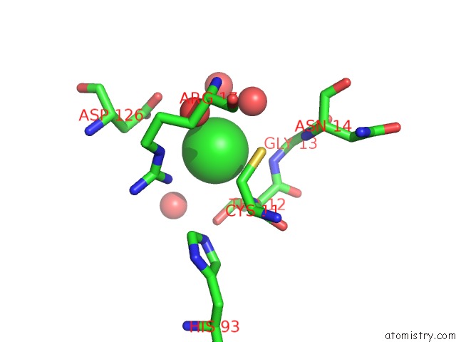
Mono view
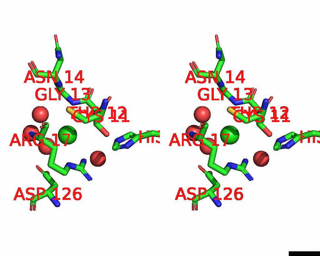
Stereo pair view
Mono view
Stereo pair view
A full contact list of Chlorine with other atoms in the Cl binding
site number 1 of Crystal Structure of Mycobacterium Tuberculosis Low Molecular Protein Tyrosine Phosphatase (Mptpa) at 1.9A Resolution within 5.0Å range:
|
Reference:
C.Madhurantakam,
E.Rajakumara,
P.A.Mazumdar,
B.Saha,
D.Mitra,
H.G.Wiker,
R.Sankaranarayanan,
A.K.Das.
Crystal Structure of Low-Molecular-Weight Protein Tyrosine Phosphatase From Mycobacterium Tuberculosis at 1.9-A Resolution J.Bacteriol. V. 187 2175 2005.
ISSN: ISSN 0021-9193
PubMed: 15743966
DOI: 10.1128/JB.187.6.2175-2181.2005
Page generated: Thu Jul 10 19:40:43 2025
ISSN: ISSN 0021-9193
PubMed: 15743966
DOI: 10.1128/JB.187.6.2175-2181.2005
Last articles
K in 5AERK in 5A4J
K in 5A8K
K in 5AEO
K in 5A9F
K in 5A71
K in 5A5G
K in 5A2D
K in 5A3S
K in 5A1I