Chlorine »
PDB 1uum-1v8f »
1v6p »
Chlorine in PDB 1v6p: Crystal Structure of Cobrotoxin
Protein crystallography data
The structure of Crystal Structure of Cobrotoxin, PDB code: 1v6p
was solved by
X.Lou,
X.Tu,
J.Wang,
M.Teng,
L.Niu,
Q.Liu,
Q.Huang,
Q.Hao,
with X-Ray Crystallography technique. A brief refinement statistics is given in the table below:
| Resolution Low / High (Å) | 10.00 / 0.87 |
| Space group | C 2 2 21 |
| Cell size a, b, c (Å), α, β, γ (°) | 46.808, 47.287, 90.018, 90.00, 90.00, 90.00 |
| R / Rfree (%) | 11.9 / 15.3 |
Other elements in 1v6p:
The structure of Crystal Structure of Cobrotoxin also contains other interesting chemical elements:
| Copper | (Cu) | 18 atoms |
| Sodium | (Na) | 4 atoms |
Chlorine Binding Sites:
The binding sites of Chlorine atom in the Crystal Structure of Cobrotoxin
(pdb code 1v6p). This binding sites where shown within
5.0 Angstroms radius around Chlorine atom.
In total only one binding site of Chlorine was determined in the Crystal Structure of Cobrotoxin, PDB code: 1v6p:
In total only one binding site of Chlorine was determined in the Crystal Structure of Cobrotoxin, PDB code: 1v6p:
Chlorine binding site 1 out of 1 in 1v6p
Go back to
Chlorine binding site 1 out
of 1 in the Crystal Structure of Cobrotoxin
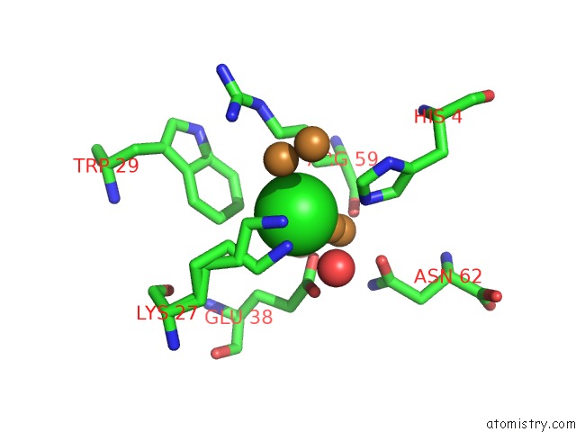
Mono view
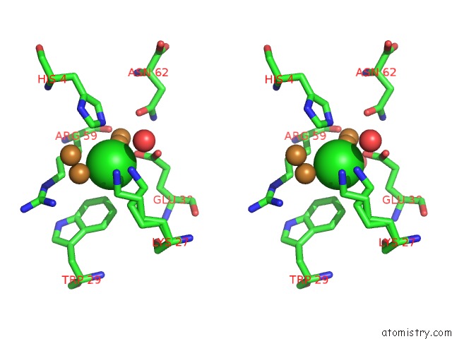
Stereo pair view
Mono view
Stereo pair view
A full contact list of Chlorine with other atoms in the Cl binding
site number 1 of Crystal Structure of Cobrotoxin within 5.0Å range:
|
Reference:
X.Lou,
Q.Liu,
X.Tu,
J.Wang,
M.Teng,
L.Niu,
D.J.Schuller,
Q.Huang,
Q.Hao.
The Atomic Resolution Crystal Structure of Atratoxin Determined By Single Wavelength Anomalous Diffraction Phasing J.Biol.Chem. V. 279 39094 2004.
ISSN: ISSN 0021-9258
PubMed: 15252034
DOI: 10.1074/JBC.M403863200
Page generated: Thu Jul 10 19:50:17 2025
ISSN: ISSN 0021-9258
PubMed: 15252034
DOI: 10.1074/JBC.M403863200
Last articles
Mn in 9LJUMn in 9LJW
Mn in 9LJS
Mn in 9LJR
Mn in 9LJT
Mn in 9LJV
Mg in 9UA2
Mg in 9R96
Mg in 9VM1
Mg in 9P01