Chlorine »
PDB 1wvu-1xkk »
1xe3 »
Chlorine in PDB 1xe3: Crystal Structure of Purine Nucleoside Phosphorylase Deod From Bacillus Anthracis
Enzymatic activity of Crystal Structure of Purine Nucleoside Phosphorylase Deod From Bacillus Anthracis
All present enzymatic activity of Crystal Structure of Purine Nucleoside Phosphorylase Deod From Bacillus Anthracis:
2.4.2.1;
2.4.2.1;
Protein crystallography data
The structure of Crystal Structure of Purine Nucleoside Phosphorylase Deod From Bacillus Anthracis, PDB code: 1xe3
was solved by
R.Grenha,
V.M.Levdikov,
M.Fogg,
E.V.Blagova,
J.A.Brannigan,
A.J.Wilkinson,
K.S.Wilson,
Structural Proteomics In Europe(Spine),
with X-Ray Crystallography technique. A brief refinement statistics is given in the table below:
| Resolution Low / High (Å) | 20.00 / 2.24 |
| Space group | P 21 21 21 |
| Cell size a, b, c (Å), α, β, γ (°) | 63.861, 128.257, 223.565, 90.00, 90.00, 90.00 |
| R / Rfree (%) | 18.1 / 23.5 |
Chlorine Binding Sites:
The binding sites of Chlorine atom in the Crystal Structure of Purine Nucleoside Phosphorylase Deod From Bacillus Anthracis
(pdb code 1xe3). This binding sites where shown within
5.0 Angstroms radius around Chlorine atom.
In total 6 binding sites of Chlorine where determined in the Crystal Structure of Purine Nucleoside Phosphorylase Deod From Bacillus Anthracis, PDB code: 1xe3:
Jump to Chlorine binding site number: 1; 2; 3; 4; 5; 6;
In total 6 binding sites of Chlorine where determined in the Crystal Structure of Purine Nucleoside Phosphorylase Deod From Bacillus Anthracis, PDB code: 1xe3:
Jump to Chlorine binding site number: 1; 2; 3; 4; 5; 6;
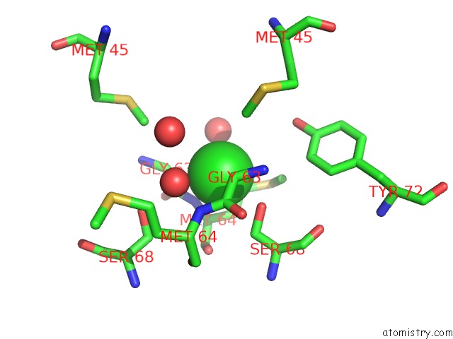
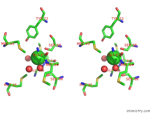
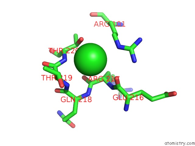
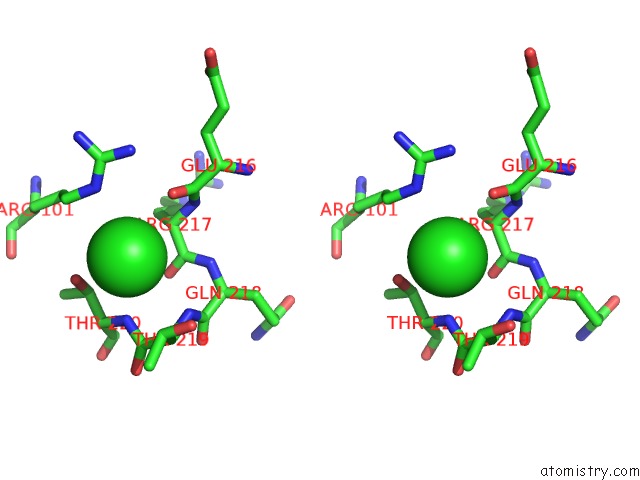
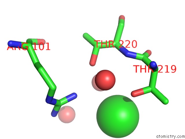
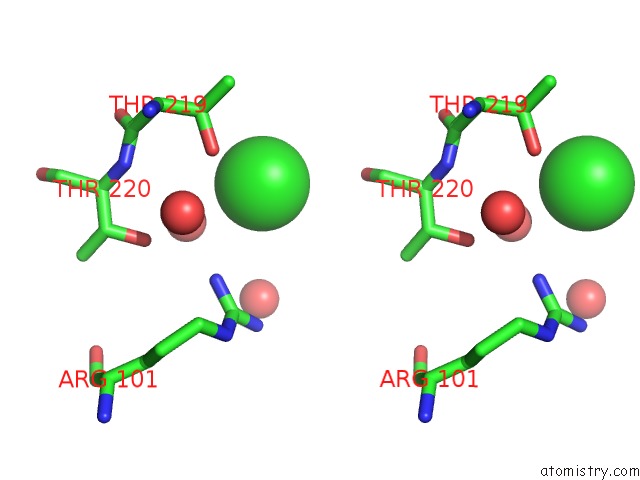
Chlorine binding site 1 out of 6 in 1xe3
Go back to
Chlorine binding site 1 out
of 6 in the Crystal Structure of Purine Nucleoside Phosphorylase Deod From Bacillus Anthracis
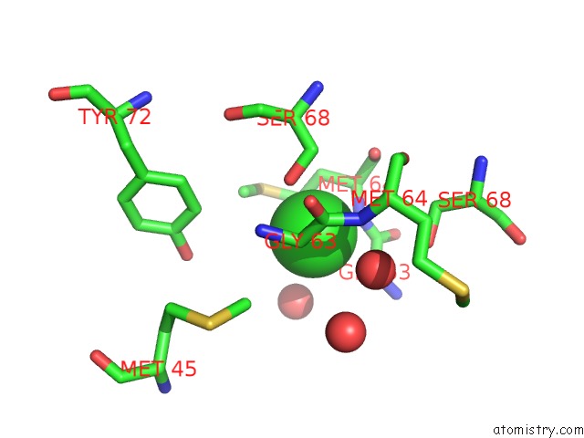
Mono view
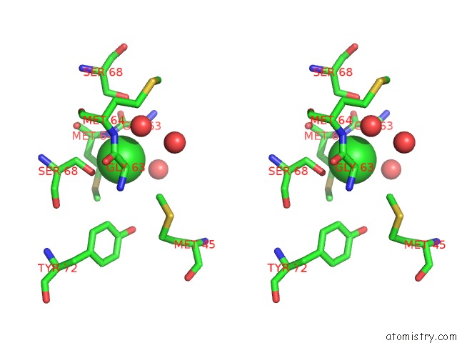
Stereo pair view
Mono view
Stereo pair view
A full contact list of Chlorine with other atoms in the Cl binding
site number 1 of Crystal Structure of Purine Nucleoside Phosphorylase Deod From Bacillus Anthracis within 5.0Å range:
|
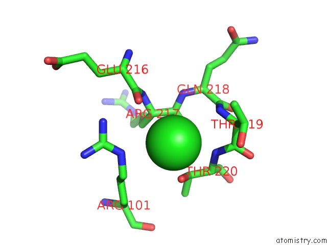
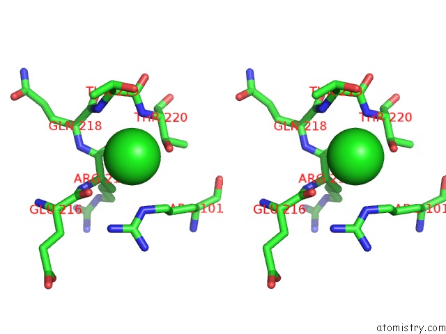
Chlorine binding site 2 out of 6 in 1xe3
Go back to
Chlorine binding site 2 out
of 6 in the Crystal Structure of Purine Nucleoside Phosphorylase Deod From Bacillus Anthracis
Mono view
Stereo pair view
Mono view
Stereo pair view
A full contact list of Chlorine with other atoms in the Cl binding
site number 2 of Crystal Structure of Purine Nucleoside Phosphorylase Deod From Bacillus Anthracis within 5.0Å range:
|
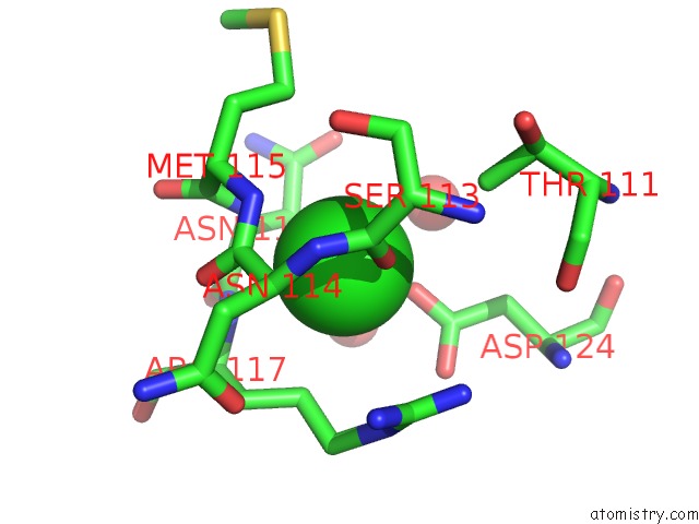
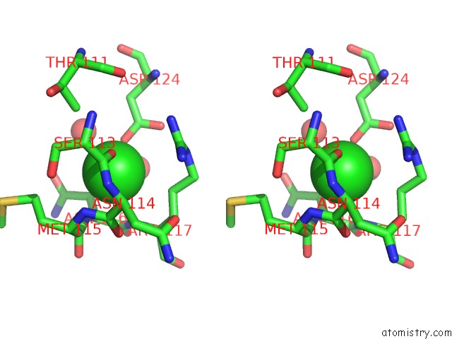
Chlorine binding site 3 out of 6 in 1xe3
Go back to
Chlorine binding site 3 out
of 6 in the Crystal Structure of Purine Nucleoside Phosphorylase Deod From Bacillus Anthracis
Mono view
Stereo pair view
Mono view
Stereo pair view
A full contact list of Chlorine with other atoms in the Cl binding
site number 3 of Crystal Structure of Purine Nucleoside Phosphorylase Deod From Bacillus Anthracis within 5.0Å range:
|
Chlorine binding site 4 out of 6 in 1xe3
Go back to
Chlorine binding site 4 out
of 6 in the Crystal Structure of Purine Nucleoside Phosphorylase Deod From Bacillus Anthracis
Mono view
Stereo pair view
Mono view
Stereo pair view
A full contact list of Chlorine with other atoms in the Cl binding
site number 4 of Crystal Structure of Purine Nucleoside Phosphorylase Deod From Bacillus Anthracis within 5.0Å range:
|
Chlorine binding site 5 out of 6 in 1xe3
Go back to
Chlorine binding site 5 out
of 6 in the Crystal Structure of Purine Nucleoside Phosphorylase Deod From Bacillus Anthracis
Mono view
Stereo pair view
Mono view
Stereo pair view
A full contact list of Chlorine with other atoms in the Cl binding
site number 5 of Crystal Structure of Purine Nucleoside Phosphorylase Deod From Bacillus Anthracis within 5.0Å range:
|
Chlorine binding site 6 out of 6 in 1xe3
Go back to
Chlorine binding site 6 out
of 6 in the Crystal Structure of Purine Nucleoside Phosphorylase Deod From Bacillus Anthracis
Mono view
Stereo pair view
Mono view
Stereo pair view
A full contact list of Chlorine with other atoms in the Cl binding
site number 6 of Crystal Structure of Purine Nucleoside Phosphorylase Deod From Bacillus Anthracis within 5.0Å range:
|
Reference:
R.Grenha,
V.M.Levdikov,
M.J.Fogg,
E.V.Blagova,
J.A.Brannigan,
A.J.Wilkinson,
K.S.Wilson.
Structure of Purine Nucleoside Phosphorylase (Deod) From Bacillus Anthracis. Acta Crystallogr.,Sect.F V. 61 459 2005.
ISSN: ESSN 1744-3091
PubMed: 16511068
DOI: 10.1107/S174430910501095X
Page generated: Thu Jul 10 20:25:11 2025
ISSN: ESSN 1744-3091
PubMed: 16511068
DOI: 10.1107/S174430910501095X
Last articles
Ni in 4NC7Ni in 4MV2
Ni in 4MSN
Ni in 4N00
Ni in 4MTQ
Ni in 4MSH
Ni in 4MTT
Ni in 4MTS
Ni in 4MSC
Ni in 4MSE