Chlorine »
PDB 1wvu-1xkk »
1xg0 »
Chlorine in PDB 1xg0: High Resolution Crystal Structure of Phycoerythrin 545 From the Marine Cryptophyte Rhodomonas CS24
Protein crystallography data
The structure of High Resolution Crystal Structure of Phycoerythrin 545 From the Marine Cryptophyte Rhodomonas CS24, PDB code: 1xg0
was solved by
A.B.Doust,
C.N.J.Marai,
S.J.Harrop,
K.E.Wilk,
P.M.G.Curmi,
G.D.Scholes,
with X-Ray Crystallography technique. A brief refinement statistics is given in the table below:
| Resolution Low / High (Å) | 60.86 / 0.97 |
| Space group | P 21 21 21 |
| Cell size a, b, c (Å), α, β, γ (°) | 62.980, 82.764, 89.477, 90.00, 90.00, 90.00 |
| R / Rfree (%) | 10.7 / 12.6 |
Other elements in 1xg0:
The structure of High Resolution Crystal Structure of Phycoerythrin 545 From the Marine Cryptophyte Rhodomonas CS24 also contains other interesting chemical elements:
| Magnesium | (Mg) | 2 atoms |
Chlorine Binding Sites:
The binding sites of Chlorine atom in the High Resolution Crystal Structure of Phycoerythrin 545 From the Marine Cryptophyte Rhodomonas CS24
(pdb code 1xg0). This binding sites where shown within
5.0 Angstroms radius around Chlorine atom.
In total only one binding site of Chlorine was determined in the High Resolution Crystal Structure of Phycoerythrin 545 From the Marine Cryptophyte Rhodomonas CS24, PDB code: 1xg0:
In total only one binding site of Chlorine was determined in the High Resolution Crystal Structure of Phycoerythrin 545 From the Marine Cryptophyte Rhodomonas CS24, PDB code: 1xg0:
Chlorine binding site 1 out of 1 in 1xg0
Go back to
Chlorine binding site 1 out
of 1 in the High Resolution Crystal Structure of Phycoerythrin 545 From the Marine Cryptophyte Rhodomonas CS24
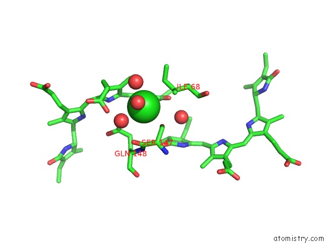
Mono view
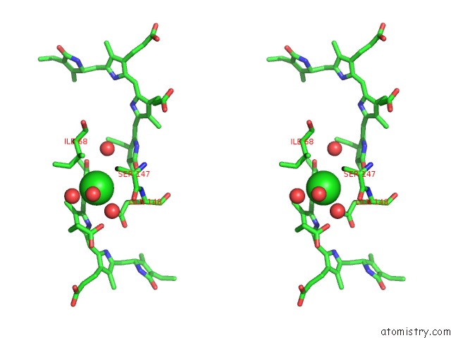
Stereo pair view
Mono view
Stereo pair view
A full contact list of Chlorine with other atoms in the Cl binding
site number 1 of High Resolution Crystal Structure of Phycoerythrin 545 From the Marine Cryptophyte Rhodomonas CS24 within 5.0Å range:
|
Reference:
A.B.Doust,
C.N.J.Marai,
S.J.Harrop,
K.E.Wilk,
P.M.G.Curmi,
G.D.Scholes.
Developing A Structure-Function Model For the Cryptophyte Phycoerythrin 545 Using Ultrahigh Resolution Crystallography and Ultrafast Laser Spectroscopy J.Mol.Biol. V. 344 135 2004.
ISSN: ISSN 0022-2836
PubMed: 15504407
DOI: 10.1016/J.JMB.2004.09.044
Page generated: Thu Jul 10 20:25:28 2025
ISSN: ISSN 0022-2836
PubMed: 15504407
DOI: 10.1016/J.JMB.2004.09.044
Last articles
Mg in 5OCGMg in 5OC0
Mg in 5OBY
Mg in 5OAT
Mg in 5OBW
Mg in 5OBU
Mg in 5OBJ
Mg in 5O7X
Mg in 5OBR
Mg in 5OAW