Chlorine »
PDB 1xkn-1y6q »
1xv5 »
Chlorine in PDB 1xv5: Alpha-Glucosyltransferase (Agt) in Complex with Udp
Enzymatic activity of Alpha-Glucosyltransferase (Agt) in Complex with Udp
All present enzymatic activity of Alpha-Glucosyltransferase (Agt) in Complex with Udp:
2.4.1.26;
2.4.1.26;
Protein crystallography data
The structure of Alpha-Glucosyltransferase (Agt) in Complex with Udp, PDB code: 1xv5
was solved by
L.Lariviere,
N.Sommer,
S.Morera,
with X-Ray Crystallography technique. A brief refinement statistics is given in the table below:
| Resolution Low / High (Å) | 20.00 / 1.73 |
| Space group | P 1 21 1 |
| Cell size a, b, c (Å), α, β, γ (°) | 47.806, 68.241, 65.750, 90.00, 109.10, 90.00 |
| R / Rfree (%) | 17.5 / 20.5 |
Chlorine Binding Sites:
The binding sites of Chlorine atom in the Alpha-Glucosyltransferase (Agt) in Complex with Udp
(pdb code 1xv5). This binding sites where shown within
5.0 Angstroms radius around Chlorine atom.
In total only one binding site of Chlorine was determined in the Alpha-Glucosyltransferase (Agt) in Complex with Udp, PDB code: 1xv5:
In total only one binding site of Chlorine was determined in the Alpha-Glucosyltransferase (Agt) in Complex with Udp, PDB code: 1xv5:
Chlorine binding site 1 out of 1 in 1xv5
Go back to
Chlorine binding site 1 out
of 1 in the Alpha-Glucosyltransferase (Agt) in Complex with Udp
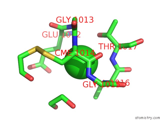
Mono view
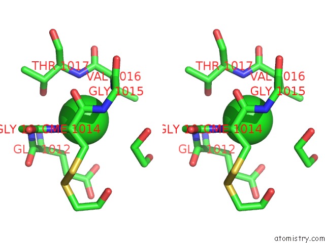
Stereo pair view
Mono view
Stereo pair view
A full contact list of Chlorine with other atoms in the Cl binding
site number 1 of Alpha-Glucosyltransferase (Agt) in Complex with Udp within 5.0Å range:
|
Reference:
L.Lariviere,
N.Sommer,
S.Morera.
Structural Evidence of A Passive Base-Flipping Mechanism For Agt, An Unusual Gt-B Glycosyltransferase. J.Mol.Biol. V. 352 139 2005.
ISSN: ISSN 0022-2836
PubMed: 16081100
DOI: 10.1016/J.JMB.2005.07.007
Page generated: Thu Jul 10 20:29:33 2025
ISSN: ISSN 0022-2836
PubMed: 16081100
DOI: 10.1016/J.JMB.2005.07.007
Last articles
Hg in 6NWHHg in 6RH4
Hg in 6RG5
Hg in 6RFH
Hg in 6PII
Hg in 6LNH
Hg in 6NJX
Hg in 6KR6
Hg in 6IQV
Hg in 6L8O