Chlorine »
PDB 1y88-1ym9 »
1yd6 »
Chlorine in PDB 1yd6: Crystal Structure of the Giy-Yig N-Terminal Endonuclease Domain of Uvrc From Bacillus Caldotenax
Protein crystallography data
The structure of Crystal Structure of the Giy-Yig N-Terminal Endonuclease Domain of Uvrc From Bacillus Caldotenax, PDB code: 1yd6
was solved by
J.J.Truglio,
B.Rhau,
D.L.Croteau,
L.Wang,
M.Skorvaga,
E.Karakas,
M.J.Dellavecchia,
H.Wang,
B.Van Houten,
C.Kisker,
with X-Ray Crystallography technique. A brief refinement statistics is given in the table below:
| Resolution Low / High (Å) | 15.00 / 2.00 |
| Space group | C 1 2 1 |
| Cell size a, b, c (Å), α, β, γ (°) | 86.487, 86.660, 67.862, 90.00, 120.16, 90.00 |
| R / Rfree (%) | 20.2 / 25.2 |
Chlorine Binding Sites:
The binding sites of Chlorine atom in the Crystal Structure of the Giy-Yig N-Terminal Endonuclease Domain of Uvrc From Bacillus Caldotenax
(pdb code 1yd6). This binding sites where shown within
5.0 Angstroms radius around Chlorine atom.
In total 3 binding sites of Chlorine where determined in the Crystal Structure of the Giy-Yig N-Terminal Endonuclease Domain of Uvrc From Bacillus Caldotenax, PDB code: 1yd6:
Jump to Chlorine binding site number: 1; 2; 3;
In total 3 binding sites of Chlorine where determined in the Crystal Structure of the Giy-Yig N-Terminal Endonuclease Domain of Uvrc From Bacillus Caldotenax, PDB code: 1yd6:
Jump to Chlorine binding site number: 1; 2; 3;
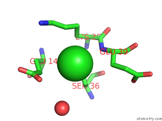
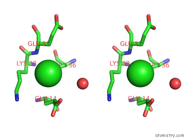
Chlorine binding site 1 out of 3 in 1yd6
Go back to
Chlorine binding site 1 out
of 3 in the Crystal Structure of the Giy-Yig N-Terminal Endonuclease Domain of Uvrc From Bacillus Caldotenax
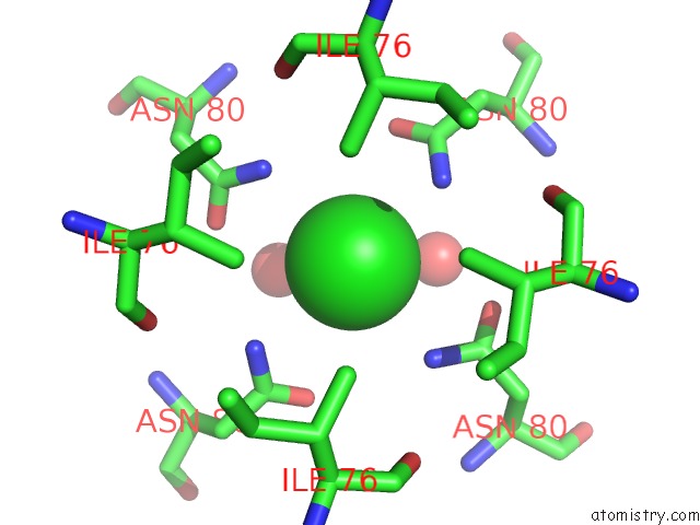
Mono view
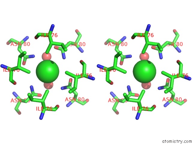
Stereo pair view
Mono view
Stereo pair view
A full contact list of Chlorine with other atoms in the Cl binding
site number 1 of Crystal Structure of the Giy-Yig N-Terminal Endonuclease Domain of Uvrc From Bacillus Caldotenax within 5.0Å range:
|
Chlorine binding site 2 out of 3 in 1yd6
Go back to
Chlorine binding site 2 out
of 3 in the Crystal Structure of the Giy-Yig N-Terminal Endonuclease Domain of Uvrc From Bacillus Caldotenax
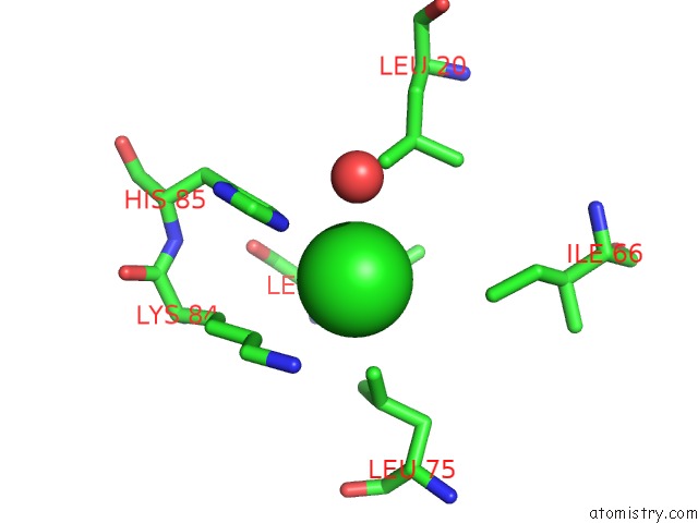
Mono view
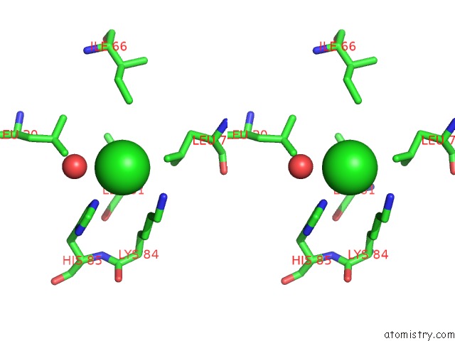
Stereo pair view
Mono view
Stereo pair view
A full contact list of Chlorine with other atoms in the Cl binding
site number 2 of Crystal Structure of the Giy-Yig N-Terminal Endonuclease Domain of Uvrc From Bacillus Caldotenax within 5.0Å range:
|
Chlorine binding site 3 out of 3 in 1yd6
Go back to
Chlorine binding site 3 out
of 3 in the Crystal Structure of the Giy-Yig N-Terminal Endonuclease Domain of Uvrc From Bacillus Caldotenax
Mono view
Stereo pair view
Mono view
Stereo pair view
A full contact list of Chlorine with other atoms in the Cl binding
site number 3 of Crystal Structure of the Giy-Yig N-Terminal Endonuclease Domain of Uvrc From Bacillus Caldotenax within 5.0Å range:
|
Reference:
J.J.Truglio,
B.Rhau,
D.L.Croteau,
L.Wang,
M.Skorvaga,
E.Karakas,
M.J.Dellavecchia,
H.Wang,
B.Van Houten,
C.Kisker.
Structural Insights Into the First Incision Reaction During Nucleotide Excision Repair Embo J. V. 24 885 2005.
ISSN: ISSN 0261-4189
PubMed: 15692561
DOI: 10.1038/SJ.EMBOJ.7600568
Page generated: Thu Jul 10 20:33:39 2025
ISSN: ISSN 0261-4189
PubMed: 15692561
DOI: 10.1038/SJ.EMBOJ.7600568
Last articles
Mg in 3HWXMg in 3HWO
Mg in 3HWW
Mg in 3HWT
Mg in 3HW5
Mg in 3HWS
Mg in 3HQP
Mg in 3HW8
Mg in 3HVR
Mg in 3HW4