Chlorine »
PDB 233l-2a72 »
2a2c »
Chlorine in PDB 2a2c: X-Ray Structure of Human N-Acetyl Galactosamine Kinase Complexed with Mg-Adp and N-Acetyl Galactosamine 1-Phosphate
Protein crystallography data
The structure of X-Ray Structure of Human N-Acetyl Galactosamine Kinase Complexed with Mg-Adp and N-Acetyl Galactosamine 1-Phosphate, PDB code: 2a2c
was solved by
J.B.Thoden,
H.M.Holden,
with X-Ray Crystallography technique. A brief refinement statistics is given in the table below:
| Resolution Low / High (Å) | 50.00 / 1.65 |
| Space group | P 65 |
| Cell size a, b, c (Å), α, β, γ (°) | 123.800, 123.800, 60.100, 90.00, 90.00, 120.00 |
| R / Rfree (%) | n/a / n/a |
Other elements in 2a2c:
The structure of X-Ray Structure of Human N-Acetyl Galactosamine Kinase Complexed with Mg-Adp and N-Acetyl Galactosamine 1-Phosphate also contains other interesting chemical elements:
| Magnesium | (Mg) | 1 atom |
| Sodium | (Na) | 1 atom |
Chlorine Binding Sites:
The binding sites of Chlorine atom in the X-Ray Structure of Human N-Acetyl Galactosamine Kinase Complexed with Mg-Adp and N-Acetyl Galactosamine 1-Phosphate
(pdb code 2a2c). This binding sites where shown within
5.0 Angstroms radius around Chlorine atom.
In total only one binding site of Chlorine was determined in the X-Ray Structure of Human N-Acetyl Galactosamine Kinase Complexed with Mg-Adp and N-Acetyl Galactosamine 1-Phosphate, PDB code: 2a2c:
In total only one binding site of Chlorine was determined in the X-Ray Structure of Human N-Acetyl Galactosamine Kinase Complexed with Mg-Adp and N-Acetyl Galactosamine 1-Phosphate, PDB code: 2a2c:
Chlorine binding site 1 out of 1 in 2a2c
Go back to
Chlorine binding site 1 out
of 1 in the X-Ray Structure of Human N-Acetyl Galactosamine Kinase Complexed with Mg-Adp and N-Acetyl Galactosamine 1-Phosphate
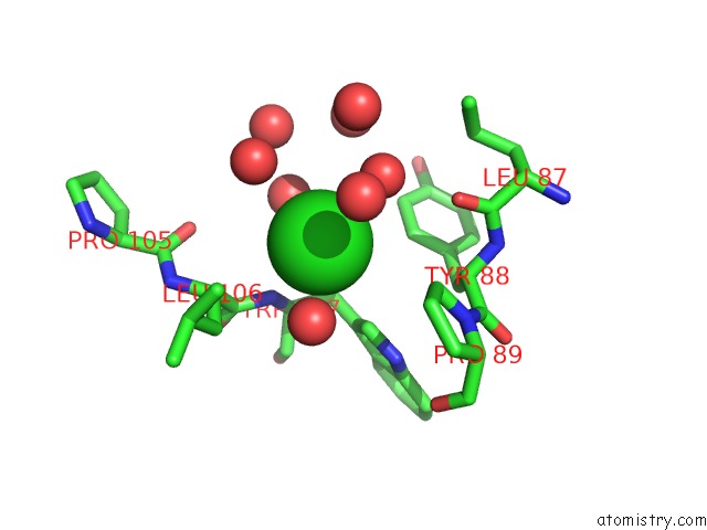
Mono view
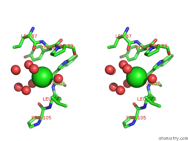
Stereo pair view
Mono view
Stereo pair view
A full contact list of Chlorine with other atoms in the Cl binding
site number 1 of X-Ray Structure of Human N-Acetyl Galactosamine Kinase Complexed with Mg-Adp and N-Acetyl Galactosamine 1-Phosphate within 5.0Å range:
|
Reference:
J.B.Thoden,
H.M.Holden.
The Molecular Architecture of Human N-Acetylgalactosamine Kinase. J.Biol.Chem. V. 280 32784 2005.
ISSN: ISSN 0021-9258
PubMed: 16006554
DOI: 10.1074/JBC.M505730200
Page generated: Thu Jul 10 21:03:55 2025
ISSN: ISSN 0021-9258
PubMed: 16006554
DOI: 10.1074/JBC.M505730200
Last articles
I in 6WXMI in 6X42
I in 6X2D
I in 6WYQ
I in 6WOK
I in 6WNY
I in 6W9D
I in 6WC8
I in 6WE7
I in 6W35