Chlorine »
PDB 2a7d-2aj4 »
2aa0 »
Chlorine in PDB 2aa0: Crystal Structure of T. Gondii Adenosine Kinase Complexed with 6-Methylmercaptopurine Riboside
Enzymatic activity of Crystal Structure of T. Gondii Adenosine Kinase Complexed with 6-Methylmercaptopurine Riboside
All present enzymatic activity of Crystal Structure of T. Gondii Adenosine Kinase Complexed with 6-Methylmercaptopurine Riboside:
2.7.1.20;
2.7.1.20;
Protein crystallography data
The structure of Crystal Structure of T. Gondii Adenosine Kinase Complexed with 6-Methylmercaptopurine Riboside, PDB code: 2aa0
was solved by
Y.Zhang,
M.H.El Kouni,
S.E.Ealick,
with X-Ray Crystallography technique. A brief refinement statistics is given in the table below:
| Resolution Low / High (Å) | 27.45 / 1.75 |
| Space group | P 21 21 21 |
| Cell size a, b, c (Å), α, β, γ (°) | 60.348, 61.701, 91.958, 90.00, 90.00, 90.00 |
| R / Rfree (%) | 20.4 / 24 |
Other elements in 2aa0:
The structure of Crystal Structure of T. Gondii Adenosine Kinase Complexed with 6-Methylmercaptopurine Riboside also contains other interesting chemical elements:
| Sodium | (Na) | 2 atoms |
Chlorine Binding Sites:
The binding sites of Chlorine atom in the Crystal Structure of T. Gondii Adenosine Kinase Complexed with 6-Methylmercaptopurine Riboside
(pdb code 2aa0). This binding sites where shown within
5.0 Angstroms radius around Chlorine atom.
In total only one binding site of Chlorine was determined in the Crystal Structure of T. Gondii Adenosine Kinase Complexed with 6-Methylmercaptopurine Riboside, PDB code: 2aa0:
In total only one binding site of Chlorine was determined in the Crystal Structure of T. Gondii Adenosine Kinase Complexed with 6-Methylmercaptopurine Riboside, PDB code: 2aa0:
Chlorine binding site 1 out of 1 in 2aa0
Go back to
Chlorine binding site 1 out
of 1 in the Crystal Structure of T. Gondii Adenosine Kinase Complexed with 6-Methylmercaptopurine Riboside
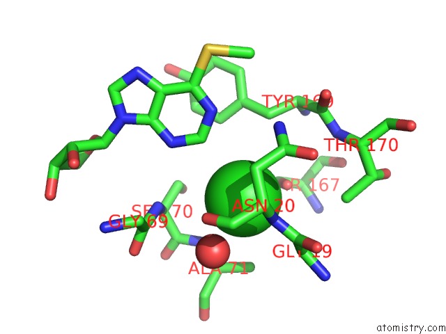
Mono view
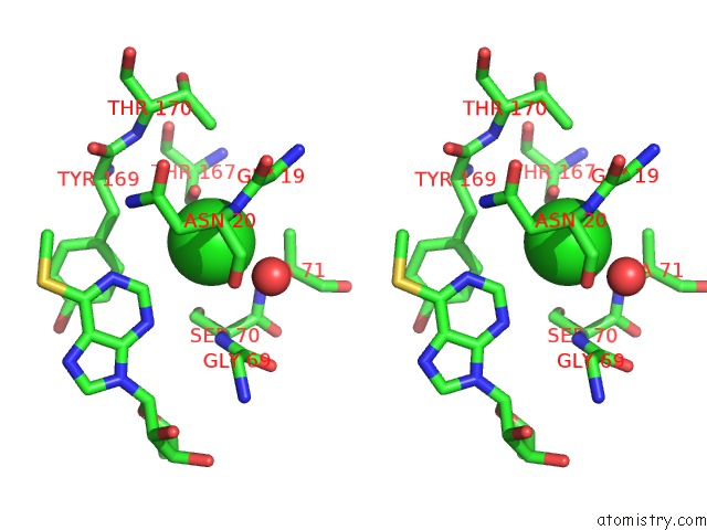
Stereo pair view
Mono view
Stereo pair view
A full contact list of Chlorine with other atoms in the Cl binding
site number 1 of Crystal Structure of T. Gondii Adenosine Kinase Complexed with 6-Methylmercaptopurine Riboside within 5.0Å range:
|
Reference:
Y.Zhang,
M.H.El Kouni,
S.E.Ealick.
Substrate Analogs Induce An Intermediate Conformational Change in Toxoplasma Gondii Adenosine Kinase Acta Crystallogr.,Sect.D V. 63 126 2007.
ISSN: ISSN 0907-4449
PubMed: 17242506
DOI: 10.1107/S0907444906043654
Page generated: Thu Jul 10 21:06:44 2025
ISSN: ISSN 0907-4449
PubMed: 17242506
DOI: 10.1107/S0907444906043654
Last articles
Fe in 2FRVFe in 2G6N
Fe in 2G6M
Fe in 2G6L
Fe in 2G6K
Fe in 2FYN
Fe in 2G6J
Fe in 2G6I
Fe in 2G6H
Fe in 2G5G