Chlorine »
PDB 2bdg-2bl9 »
2bl9 »
Chlorine in PDB 2bl9: X-Ray Crystal Structure of Plasmodium Vivax Dihydrofolate Reductase in Complex with Pyrimethamine and Its Derivative
Enzymatic activity of X-Ray Crystal Structure of Plasmodium Vivax Dihydrofolate Reductase in Complex with Pyrimethamine and Its Derivative
All present enzymatic activity of X-Ray Crystal Structure of Plasmodium Vivax Dihydrofolate Reductase in Complex with Pyrimethamine and Its Derivative:
1.5.1.3;
1.5.1.3;
Protein crystallography data
The structure of X-Ray Crystal Structure of Plasmodium Vivax Dihydrofolate Reductase in Complex with Pyrimethamine and Its Derivative, PDB code: 2bl9
was solved by
P.Kongsaeree,
P.Khongsuk,
U.Leartsakulpanich,
P.Chitnumsub,
B.Tarnchompoo,
M.D.Walkinshaw,
Y.Yuthavong,
with X-Ray Crystallography technique. A brief refinement statistics is given in the table below:
| Resolution Low / High (Å) | 30.47 / 1.9 |
| Space group | C 1 2 1 |
| Cell size a, b, c (Å), α, β, γ (°) | 134.280, 55.740, 45.720, 90.00, 107.09, 90.00 |
| R / Rfree (%) | 20.8 / 26 |
Chlorine Binding Sites:
The binding sites of Chlorine atom in the X-Ray Crystal Structure of Plasmodium Vivax Dihydrofolate Reductase in Complex with Pyrimethamine and Its Derivative
(pdb code 2bl9). This binding sites where shown within
5.0 Angstroms radius around Chlorine atom.
In total only one binding site of Chlorine was determined in the X-Ray Crystal Structure of Plasmodium Vivax Dihydrofolate Reductase in Complex with Pyrimethamine and Its Derivative, PDB code: 2bl9:
In total only one binding site of Chlorine was determined in the X-Ray Crystal Structure of Plasmodium Vivax Dihydrofolate Reductase in Complex with Pyrimethamine and Its Derivative, PDB code: 2bl9:
Chlorine binding site 1 out of 1 in 2bl9
Go back to
Chlorine binding site 1 out
of 1 in the X-Ray Crystal Structure of Plasmodium Vivax Dihydrofolate Reductase in Complex with Pyrimethamine and Its Derivative
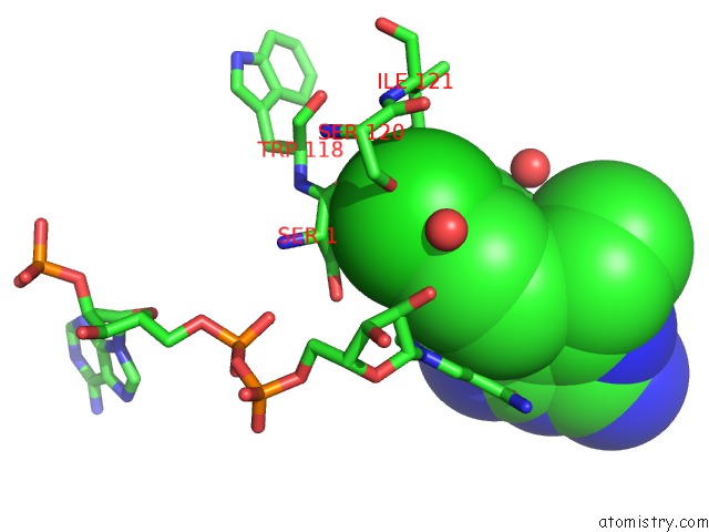
Mono view
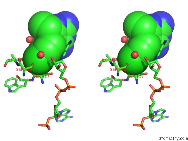
Stereo pair view
Mono view
Stereo pair view
A full contact list of Chlorine with other atoms in the Cl binding
site number 1 of X-Ray Crystal Structure of Plasmodium Vivax Dihydrofolate Reductase in Complex with Pyrimethamine and Its Derivative within 5.0Å range:
|
Reference:
P.Kongsaeree,
P.Khongsuk,
U.Leartsakulpanich,
P.Chitnumsub,
B.Tarnchompoo,
M.D.Walkinshaw,
Y.Yuthavong.
Crystal Structure of Dihydrofolate Reductase From Plasmodium Vivax: Pyrimethamine Displacement Linked with Mutation-Induced Resistance. Proc.Natl.Acad.Sci.Usa V. 102 13046 2005.
ISSN: ISSN 0027-8424
PubMed: 16135570
DOI: 10.1073/PNAS.0501747102
Page generated: Thu Jul 10 21:28:08 2025
ISSN: ISSN 0027-8424
PubMed: 16135570
DOI: 10.1073/PNAS.0501747102
Last articles
Mg in 4JI7Mg in 4JI6
Mg in 4JJS
Mg in 4JJ2
Mg in 4JIW
Mg in 4JIV
Mg in 4JIB
Mg in 4JI4
Mg in 4JI5
Mg in 4JI1