Chlorine »
PDB 2bwx-2c5u »
2bzh »
Chlorine in PDB 2bzh: Crystal Structure of the Human PIM1 in Complex with A Ruthenium Organometallic Ligand RU1
Enzymatic activity of Crystal Structure of the Human PIM1 in Complex with A Ruthenium Organometallic Ligand RU1
All present enzymatic activity of Crystal Structure of the Human PIM1 in Complex with A Ruthenium Organometallic Ligand RU1:
2.7.1.37;
2.7.1.37;
Protein crystallography data
The structure of Crystal Structure of the Human PIM1 in Complex with A Ruthenium Organometallic Ligand RU1, PDB code: 2bzh
was solved by
J.E.Debreczeni,
A.Bullock,
S.Knapp,
F.Von Delft,
M.Sundstrom,
C.Arrowsmith,
J.Weigelt,
A.Edwards,
with X-Ray Crystallography technique. A brief refinement statistics is given in the table below:
| Resolution Low / High (Å) | 40.00 / 1.90 |
| Space group | P 65 |
| Cell size a, b, c (Å), α, β, γ (°) | 97.529, 97.529, 80.726, 90.00, 90.00, 120.00 |
| R / Rfree (%) | 15.7 / 19 |
Other elements in 2bzh:
The structure of Crystal Structure of the Human PIM1 in Complex with A Ruthenium Organometallic Ligand RU1 also contains other interesting chemical elements:
| Ruthenium | (Ru) | 1 atom |
Chlorine Binding Sites:
The binding sites of Chlorine atom in the Crystal Structure of the Human PIM1 in Complex with A Ruthenium Organometallic Ligand RU1
(pdb code 2bzh). This binding sites where shown within
5.0 Angstroms radius around Chlorine atom.
In total only one binding site of Chlorine was determined in the Crystal Structure of the Human PIM1 in Complex with A Ruthenium Organometallic Ligand RU1, PDB code: 2bzh:
In total only one binding site of Chlorine was determined in the Crystal Structure of the Human PIM1 in Complex with A Ruthenium Organometallic Ligand RU1, PDB code: 2bzh:
Chlorine binding site 1 out of 1 in 2bzh
Go back to
Chlorine binding site 1 out
of 1 in the Crystal Structure of the Human PIM1 in Complex with A Ruthenium Organometallic Ligand RU1
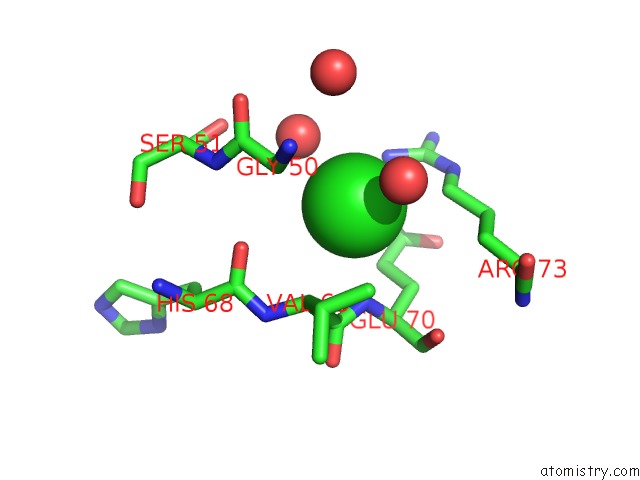
Mono view
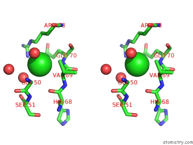
Stereo pair view
Mono view
Stereo pair view
A full contact list of Chlorine with other atoms in the Cl binding
site number 1 of Crystal Structure of the Human PIM1 in Complex with A Ruthenium Organometallic Ligand RU1 within 5.0Å range:
|
Reference:
J.E.Debreczeni,
A.Bullock,
S.Knapp,
F.Von Delft,
M.Sundstrom,
C.Arrowsmith,
J.Weigelt,
A.Edwards.
Crystal Structure of the Human PIM1 in Complex with Ruthenium Organometallic Ligands To Be Published.
Page generated: Thu Jul 10 21:34:51 2025
Last articles
Mg in 1XNGMg in 1XN0
Mg in 1XMY
Mg in 1XMV
Mg in 1XMX
Mg in 1XMI
Mg in 1XMU
Mg in 1XM6
Mg in 1XMJ
Mg in 1XM4