Chlorine »
PDB 2dxd-2esw »
2e66 »
Chlorine in PDB 2e66: Crystal Structure of CUTA1 From Pyrococcus Horikoshii OT3, Mutation D60A
Protein crystallography data
The structure of Crystal Structure of CUTA1 From Pyrococcus Horikoshii OT3, Mutation D60A, PDB code: 2e66
was solved by
B.Bagautdinov,
M.Sawano,
S.Bagautdinova,
K.Yutani,
N.Kunishima,
Rikenstructural Genomics/Proteomics Initiative (Rsgi),
with X-Ray Crystallography technique. A brief refinement statistics is given in the table below:
| Resolution Low / High (Å) | 35.88 / 2.00 |
| Space group | P 21 21 21 |
| Cell size a, b, c (Å), α, β, γ (°) | 44.191, 76.348, 103.488, 90.00, 90.00, 90.00 |
| R / Rfree (%) | 22.9 / 25.3 |
Other elements in 2e66:
The structure of Crystal Structure of CUTA1 From Pyrococcus Horikoshii OT3, Mutation D60A also contains other interesting chemical elements:
| Sodium | (Na) | 1 atom |
Chlorine Binding Sites:
The binding sites of Chlorine atom in the Crystal Structure of CUTA1 From Pyrococcus Horikoshii OT3, Mutation D60A
(pdb code 2e66). This binding sites where shown within
5.0 Angstroms radius around Chlorine atom.
In total only one binding site of Chlorine was determined in the Crystal Structure of CUTA1 From Pyrococcus Horikoshii OT3, Mutation D60A, PDB code: 2e66:
In total only one binding site of Chlorine was determined in the Crystal Structure of CUTA1 From Pyrococcus Horikoshii OT3, Mutation D60A, PDB code: 2e66:
Chlorine binding site 1 out of 1 in 2e66
Go back to
Chlorine binding site 1 out
of 1 in the Crystal Structure of CUTA1 From Pyrococcus Horikoshii OT3, Mutation D60A
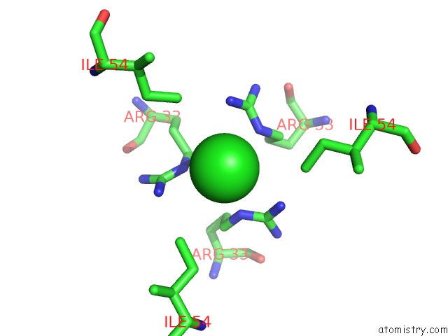
Mono view
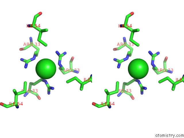
Stereo pair view
Mono view
Stereo pair view
A full contact list of Chlorine with other atoms in the Cl binding
site number 1 of Crystal Structure of CUTA1 From Pyrococcus Horikoshii OT3, Mutation D60A within 5.0Å range:
|
Reference:
B.Bagautdinov,
M.Sawano,
S.Bagautdinova,
K.Yutani,
N.Kunishima.
Structural Basis of the Hyper-Thermostability of CUTA1 To Be Published.
Page generated: Thu Jul 10 21:59:01 2025
Last articles
Mg in 6JLJMg in 6JIL
Mg in 6JKM
Mg in 6JJW
Mg in 6JJU
Mg in 6JJ9
Mg in 6JD2
Mg in 6JJ8
Mg in 6JIM
Mg in 6JD1