Chlorine »
PDB 2dxd-2esw »
2eg1 »
Chlorine in PDB 2eg1: The Crystal Structure of Pii Protein
Protein crystallography data
The structure of The Crystal Structure of Pii Protein, PDB code: 2eg1
was solved by
H.Sakai,
A.Shinkai,
Y.Kitamura,
S.Kuramitsu,
S.Yokoyama,
Riken Structuralgenomics/Proteomics Initiative (Rsgi),
with X-Ray Crystallography technique. A brief refinement statistics is given in the table below:
| Resolution Low / High (Å) | 40.52 / 1.80 |
| Space group | I 2 3 |
| Cell size a, b, c (Å), α, β, γ (°) | 81.040, 81.040, 81.040, 90.00, 90.00, 90.00 |
| R / Rfree (%) | 18.5 / 22.3 |
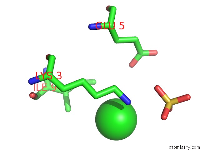
Chlorine Binding Sites:
The binding sites of Chlorine atom in the The Crystal Structure of Pii Protein
(pdb code 2eg1). This binding sites where shown within
5.0 Angstroms radius around Chlorine atom.
In total only one binding site of Chlorine was determined in the The Crystal Structure of Pii Protein, PDB code: 2eg1:
In total only one binding site of Chlorine was determined in the The Crystal Structure of Pii Protein, PDB code: 2eg1:
Chlorine binding site 1 out of 1 in 2eg1
Go back to
Chlorine binding site 1 out
of 1 in the The Crystal Structure of Pii Protein
Mono view

Stereo pair view
Mono view
Stereo pair view
A full contact list of Chlorine with other atoms in the Cl binding
site number 1 of The Crystal Structure of Pii Protein within 5.0Å range:
|
Reference:
H.Sakai,
A.Shinkai,
Y.Kitamura,
S.Kuramitsu,
S.Yokoyama.
The Crystal Structure of Pii Protein To Be Published.
Page generated: Thu Jul 10 21:59:47 2025
Last articles
Mg in 6JNXMg in 6JMG
Mg in 6JNL
Mg in 6JLV
Mg in 6JLN
Mg in 6JLL
Mg in 6JLK
Mg in 6JLM
Mg in 6JLJ
Mg in 6JIL