Chlorine »
PDB 2etd-2f81 »
2f1r »
Chlorine in PDB 2f1r: Crystal Structure of Molybdopterin-Guanine Biosynthesis Protein B (Mobb)
Protein crystallography data
The structure of Crystal Structure of Molybdopterin-Guanine Biosynthesis Protein B (Mobb), PDB code: 2f1r
was solved by
L.Damodharan,
S.Eswaramoorthy,
D.Kumaran,
S.Swaminathan,
S.K.Burley,
New York Sgx Research Center For Structuralgenomics (Nysgxrc),
with X-Ray Crystallography technique. A brief refinement statistics is given in the table below:
| Resolution Low / High (Å) | 50.00 / 2.10 |
| Space group | P 1 21 1 |
| Cell size a, b, c (Å), α, β, γ (°) | 37.980, 63.930, 66.010, 90.00, 95.30, 90.00 |
| R / Rfree (%) | 18.6 / 25.3 |
Other elements in 2f1r:
The structure of Crystal Structure of Molybdopterin-Guanine Biosynthesis Protein B (Mobb) also contains other interesting chemical elements:
| Praseodymium | (Pr) | 1 atom |
Chlorine Binding Sites:
The binding sites of Chlorine atom in the Crystal Structure of Molybdopterin-Guanine Biosynthesis Protein B (Mobb)
(pdb code 2f1r). This binding sites where shown within
5.0 Angstroms radius around Chlorine atom.
In total only one binding site of Chlorine was determined in the Crystal Structure of Molybdopterin-Guanine Biosynthesis Protein B (Mobb), PDB code: 2f1r:
In total only one binding site of Chlorine was determined in the Crystal Structure of Molybdopterin-Guanine Biosynthesis Protein B (Mobb), PDB code: 2f1r:
Chlorine binding site 1 out of 1 in 2f1r
Go back to Chlorine Binding Sites List in 2f1r
Chlorine binding site 1 out
of 1 in the Crystal Structure of Molybdopterin-Guanine Biosynthesis Protein B (Mobb)
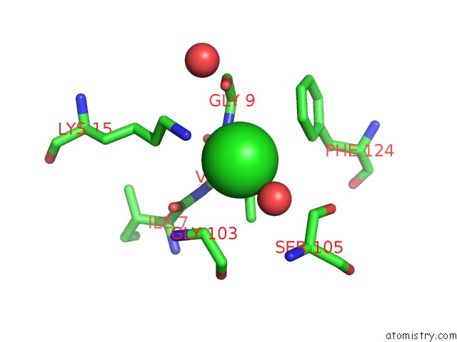
Mono view
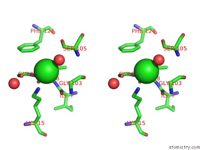
Stereo pair view
Mono view
Stereo pair view
A full contact list of Chlorine with other atoms in the Cl binding
site number 1 of Crystal Structure of Molybdopterin-Guanine Biosynthesis Protein B (Mobb) within 5.0Å range:
|
Reference:
L.Damodharan,
S.Eswaramoorthy,
D.Kumaran,
S.Swaminathan.
Crystal Structure of Molybdopterin-Guanine Dinucleotide Biosynthesis Protein B (Mobb) To Be Published.
Page generated: Sat Jul 20 06:53:00 2024
Last articles
Zn in 9MJ5Zn in 9HNW
Zn in 9G0L
Zn in 9FNE
Zn in 9DZN
Zn in 9E0I
Zn in 9D32
Zn in 9DAK
Zn in 8ZXC
Zn in 8ZUF
