Chlorine »
PDB 2etd-2f81 »
2f2s »
Chlorine in PDB 2f2s: Human Mitochondrial Acetoacetyl-Coa Thiolase
Enzymatic activity of Human Mitochondrial Acetoacetyl-Coa Thiolase
All present enzymatic activity of Human Mitochondrial Acetoacetyl-Coa Thiolase:
2.3.1.9;
2.3.1.9;
Protein crystallography data
The structure of Human Mitochondrial Acetoacetyl-Coa Thiolase, PDB code: 2f2s
was solved by
J.R.Min,
L.Dombrovski,
T.Antoshenko,
H.Wu,
P.Loppnau,
J.Weigelt,
M.Sundstrom,
C.H.Arrowsmith,
A.M.Edwards,
A.Bochkarev,
A.N.Plotnikov,
Structural Genomics Consortium (Sgc),
with X-Ray Crystallography technique. A brief refinement statistics is given in the table below:
| Resolution Low / High (Å) | 33.80 / 2.00 |
| Space group | P 1 21 1 |
| Cell size a, b, c (Å), α, β, γ (°) | 56.989, 126.640, 111.858, 90.00, 98.64, 90.00 |
| R / Rfree (%) | 19.9 / 25.7 |
Chlorine Binding Sites:
The binding sites of Chlorine atom in the Human Mitochondrial Acetoacetyl-Coa Thiolase
(pdb code 2f2s). This binding sites where shown within
5.0 Angstroms radius around Chlorine atom.
In total 3 binding sites of Chlorine where determined in the Human Mitochondrial Acetoacetyl-Coa Thiolase, PDB code: 2f2s:
Jump to Chlorine binding site number: 1; 2; 3;
In total 3 binding sites of Chlorine where determined in the Human Mitochondrial Acetoacetyl-Coa Thiolase, PDB code: 2f2s:
Jump to Chlorine binding site number: 1; 2; 3;
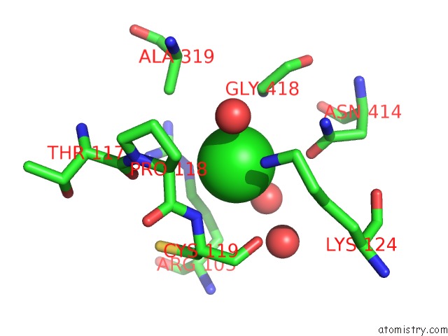
Chlorine binding site 1 out of 3 in 2f2s
Go back to
Chlorine binding site 1 out
of 3 in the Human Mitochondrial Acetoacetyl-Coa Thiolase

Mono view
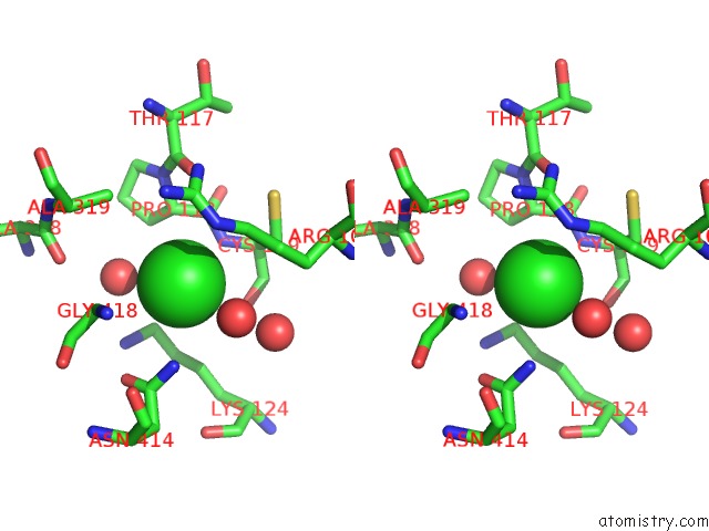
Stereo pair view
Mono view
Stereo pair view
A full contact list of Chlorine with other atoms in the Cl binding
site number 1 of Human Mitochondrial Acetoacetyl-Coa Thiolase within 5.0Å range:
|
Chlorine binding site 2 out of 3 in 2f2s
Go back to
Chlorine binding site 2 out
of 3 in the Human Mitochondrial Acetoacetyl-Coa Thiolase

Mono view

Stereo pair view
Mono view
Stereo pair view
A full contact list of Chlorine with other atoms in the Cl binding
site number 2 of Human Mitochondrial Acetoacetyl-Coa Thiolase within 5.0Å range:
|
Chlorine binding site 3 out of 3 in 2f2s
Go back to
Chlorine binding site 3 out
of 3 in the Human Mitochondrial Acetoacetyl-Coa Thiolase
Mono view

Stereo pair view
Mono view
Stereo pair view
A full contact list of Chlorine with other atoms in the Cl binding
site number 3 of Human Mitochondrial Acetoacetyl-Coa Thiolase within 5.0Å range:
|
Reference:
L.Dombrovski,
J.R.Min,
T.Antoshenko,
H.Wu,
P.Loppnau,
A.M.Edwards,
C.H.Arrowsmith,
A.Bochkarev,
A.N.Plotnikov.
The Crystal Structure of Human Mitochondrial Acetoacetyl-Coa Thiolase ACAT1. To Be Published.
Page generated: Thu Jul 10 22:02:29 2025
Last articles
Mg in 2VSCMg in 2VPN
Mg in 2VRN
Mg in 2VPQ
Mg in 2VQD
Mg in 2VQ2
Mg in 2VPR
Mg in 2VPO
Mg in 2VP0
Mg in 2VOS