Chlorine »
PDB 2fs7-2g7z »
2fvv »
Chlorine in PDB 2fvv: Human Diphosphoinositol Polyphosphate Phosphohydrolase 1
Enzymatic activity of Human Diphosphoinositol Polyphosphate Phosphohydrolase 1
All present enzymatic activity of Human Diphosphoinositol Polyphosphate Phosphohydrolase 1:
3.6.1.52;
3.6.1.52;
Protein crystallography data
The structure of Human Diphosphoinositol Polyphosphate Phosphohydrolase 1, PDB code: 2fvv
was solved by
B.M.Hallberg,
P.Kursula,
D.Ogg,
C.Arrowsmith,
H.Berglund,
A.Edwards,
M.Ehn,
S.Flodin,
S.Graslund,
M.Hammarstrom,
M.Hogbom,
L.Holmberg-Schiavone,
T.Kotenyova,
P.Nilsson-Ehle,
P.Nordlund,
T.Nyman,
J.Sagemark,
P.Stenmark,
M.Sundstrom,
A.G.Thorsell,
S.Van Den Berg,
J.Weigelt,
C.Persson,
Structural Genomics Consortium (Sgc),
with X-Ray Crystallography technique. A brief refinement statistics is given in the table below:
| Resolution Low / High (Å) | 10.00 / 1.25 |
| Space group | P 21 21 21 |
| Cell size a, b, c (Å), α, β, γ (°) | 48.653, 59.900, 61.735, 90.00, 90.00, 90.00 |
| R / Rfree (%) | 16.8 / 20.4 |
Chlorine Binding Sites:
The binding sites of Chlorine atom in the Human Diphosphoinositol Polyphosphate Phosphohydrolase 1
(pdb code 2fvv). This binding sites where shown within
5.0 Angstroms radius around Chlorine atom.
In total only one binding site of Chlorine was determined in the Human Diphosphoinositol Polyphosphate Phosphohydrolase 1, PDB code: 2fvv:
In total only one binding site of Chlorine was determined in the Human Diphosphoinositol Polyphosphate Phosphohydrolase 1, PDB code: 2fvv:
Chlorine binding site 1 out of 1 in 2fvv
Go back to
Chlorine binding site 1 out
of 1 in the Human Diphosphoinositol Polyphosphate Phosphohydrolase 1
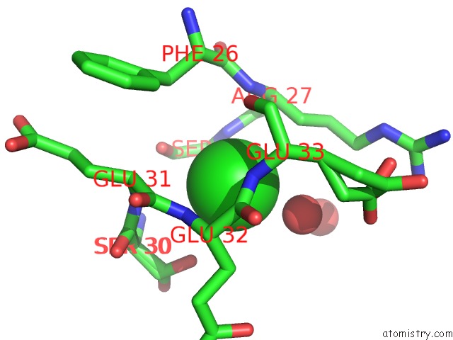
Mono view
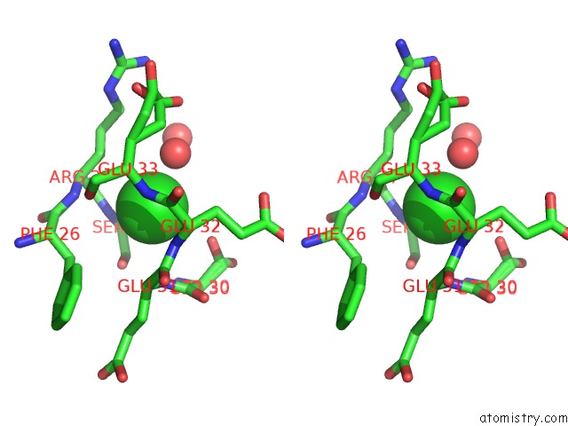
Stereo pair view
Mono view
Stereo pair view
A full contact list of Chlorine with other atoms in the Cl binding
site number 1 of Human Diphosphoinositol Polyphosphate Phosphohydrolase 1 within 5.0Å range:
|
Reference:
A.G.Thorsell,
C.Persson,
S.Graslund,
M.Hammarstrom,
R.D.Busam,
B.M.Hallberg.
Crystal Structure of Human Diphosphoinositol Phosphatase 1 Proteins V. 77 242 2009.
ISSN: ISSN 0887-3585
PubMed: 19585659
DOI: 10.1002/PROT.22489
Page generated: Thu Jul 10 22:13:16 2025
ISSN: ISSN 0887-3585
PubMed: 19585659
DOI: 10.1002/PROT.22489
Last articles
Fe in 5TH5Fe in 5T5M
Fe in 5TIA
Fe in 5TI9
Fe in 5TGS
Fe in 5TFU
Fe in 5TG0
Fe in 5TFT
Fe in 5TE8
Fe in 5TDV