Chlorine »
PDB 2fs7-2g7z »
2fyr »
Chlorine in PDB 2fyr: Crystal Structure of Norwalk Virus Protease Grown in the Presence of Aebsf
Protein crystallography data
The structure of Crystal Structure of Norwalk Virus Protease Grown in the Presence of Aebsf, PDB code: 2fyr
was solved by
C.E.Zeitler,
M.K.Estes,
B.V.Venkataram Prasad,
with X-Ray Crystallography technique. A brief refinement statistics is given in the table below:
| Resolution Low / High (Å) | 40.00 / 2.20 |
| Space group | P 65 2 2 |
| Cell size a, b, c (Å), α, β, γ (°) | 120.490, 120.490, 51.249, 90.00, 90.00, 120.00 |
| R / Rfree (%) | 21.1 / 24.6 |
Other elements in 2fyr:
The structure of Crystal Structure of Norwalk Virus Protease Grown in the Presence of Aebsf also contains other interesting chemical elements:
| Magnesium | (Mg) | 1 atom |
Chlorine Binding Sites:
The binding sites of Chlorine atom in the Crystal Structure of Norwalk Virus Protease Grown in the Presence of Aebsf
(pdb code 2fyr). This binding sites where shown within
5.0 Angstroms radius around Chlorine atom.
In total only one binding site of Chlorine was determined in the Crystal Structure of Norwalk Virus Protease Grown in the Presence of Aebsf, PDB code: 2fyr:
In total only one binding site of Chlorine was determined in the Crystal Structure of Norwalk Virus Protease Grown in the Presence of Aebsf, PDB code: 2fyr:
Chlorine binding site 1 out of 1 in 2fyr
Go back to
Chlorine binding site 1 out
of 1 in the Crystal Structure of Norwalk Virus Protease Grown in the Presence of Aebsf
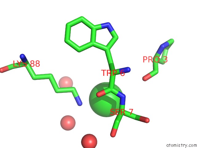
Mono view
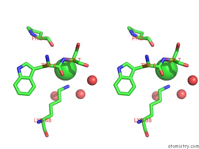
Stereo pair view
Mono view
Stereo pair view
A full contact list of Chlorine with other atoms in the Cl binding
site number 1 of Crystal Structure of Norwalk Virus Protease Grown in the Presence of Aebsf within 5.0Å range:
|
Reference:
C.E.Zeitler,
M.K.Estes,
B.V.Venkataram Prasad.
X-Ray Crystallographic Structure of the Norwalk Virus Protease at 1.5-A Resolution. J.Virol. V. 80 5050 2006.
ISSN: ISSN 0022-538X
PubMed: 16641296
DOI: 10.1128/JVI.80.10.5050-5058.2006
Page generated: Thu Jul 10 22:14:17 2025
ISSN: ISSN 0022-538X
PubMed: 16641296
DOI: 10.1128/JVI.80.10.5050-5058.2006
Last articles
Mg in 2A6EMg in 2A5Z
Mg in 2A5L
Mg in 2A5Y
Mg in 2A5J
Mg in 2A43
Mg in 2A5G
Mg in 2A5D
Mg in 2A5F
Mg in 2A42